. 2010 Sep 1;78(1):297-305.
doi: 10.1016/j.ijrobp.2009.11.008. Epub 2010 Apr 13.

Development of a micro-computed tomography-based image-guided conformal radiotherapy system for small animals

Affiliations

Development of a micro-computed tomography-based image-guided conformal radiotherapy system for small animals

Hu Zhou et al. Int J Radiat Oncol Biol Phys. .

Abstract

Purpose: To report on the physical aspects of a system in which radiotherapy functionality was added to a micro-computed tomography (microCT) scanner, to evaluate the accuracy of this instrument, and to and demonstrate the application of this technology for irradiating tumors growing within the lungs of mice.

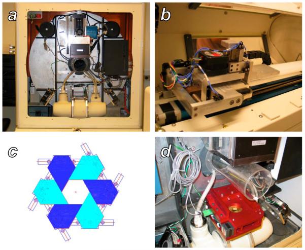
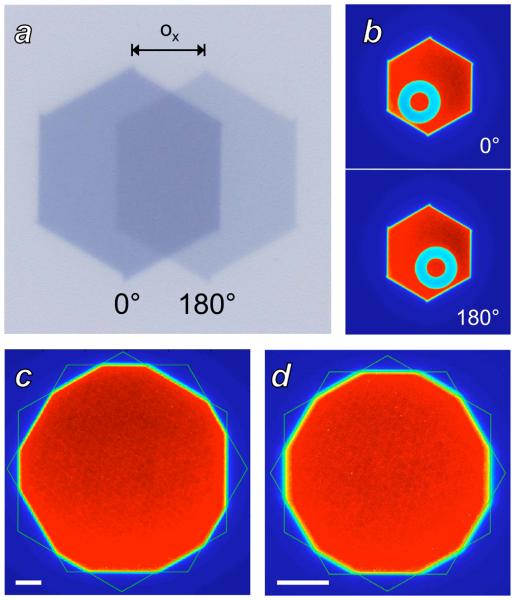
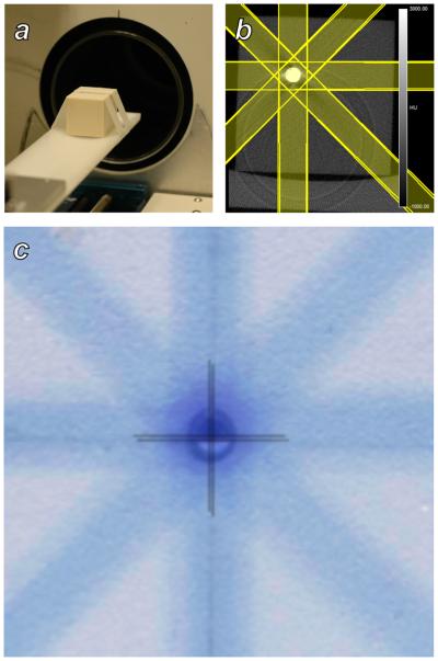
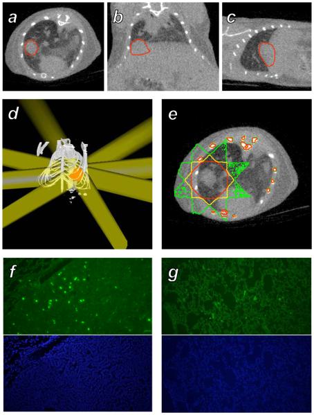
Methods and materials: A GE eXplore RS120 microCT scanner was modified by the addition of a two-dimensional subject translation stage and a variable aperture collimator. Quality assurance protocols for these devices, including measurement of translation stage positioning accuracy, collimator aperture accuracy, and collimator alignment with the X-ray beam, were devised. Use of this system for image-guided radiotherapy was assessed by irradiation of a solid water phantom as well as of two mice bearing spontaneous MYC-induced lung tumors. Radiation damage was assessed ex vivo by immunohistochemical detection of gammaH2AX foci.

Results: The positioning error of the translation stage was found to be <0.05 mm, whereas after alignment of the collimator with the X-ray axis through adjustment of its displacement and rotation, the collimator aperture error was <0.1 mm measured at isocenter. Computed tomography image-guided treatment of a solid water phantom demonstrated target localization accuracy to within 0.1 mm. Gamma-H2AX foci were detected within irradiated lung tumors in mice, with contralateral lung tissue displaying background staining.

Conclusions: Addition of radiotherapy functionality to a microCT scanner is an effective means of introducing image-guided radiation treatments into the preclinical setting. This approach has been shown to facilitate small-animal conformal radiotherapy while leveraging existing technology.


Figures

Figure 1
Figure 1
The GE RS120 microCT and the additional components necessary for small animal radiotherapy. A: A view of the scanner gantry after removing the x-ray shield and animal stage housing. The large silver disk is the rotating gantry, with the x-ray tube, H-shaped collimator mounting bar, subject bore, and detector arranged bottom to top, respectively. B: The custom two-dimensional translation stage for subject positioning, mounted on top of the existing z-axis translation stage of the scanner. C: Schematic representation of a single stage of the variable-aperture collimator, formed by six sliding blocks mounted on linear tracks. D: The final two-stage collimator apparatus after installation on the H mounting bar of the scanner gantry.
Figure 2
Figure 2
Measurement of collimator alignment with the x-ray beam axis. A: Split field irradiation. The offset ox between the images from 0° (darker) and 180° (lighter) gantry angles is used to compute the offset of the collimator from the beam axis. B: Measurement of collimator offset by imaging a fixed object. The average of the object location seen at 0° and at 180° gives the scanner isocenter, while the collimator center can be measured based on the hexagonal profile. C: Measurement of collimator offset by fitting the hexagonal profiles produced by each collimator stage, with both stages set at isocenter apertures of 20 mm. Asymmetry in the dodecagon is apparent, produced by offset between the centers of the two hexagons. The white scale bar is of length 2.5 mm at isocenter. D: Two hexagon analysis after iterative alignment of the collimator with the x-ray beam axis. The collimator apertures are set to 10 mm at isocenter to improve visual detection of any misalignment. The white scale bar is of length 2.5 mm at isocenter.
Figure 3
Figure 3
Evaluation of subject targeting accuracy using a solid water phantom. A: A phantom containing a metal sphere and radiochromic film was placed on the scanner bed and imaged with microCT. B: The microCT image of the phantom showing the metal inclusion, and the radiation treatment plan that was constructed in RT_Image. C: A superposition of a the solid water/metal sphere phantom and the adjacent irradiated film. Crosshairs identify the positions of the center of the sphere and the center of the delivered dose distribution, which agree to within 0.1 mm.
Figure 4
Figure 4
Treatment of a murine spontaneous lung tumor with the microCT radiotherapy system. A-C: A MYC-induced tumor growing in the base of the right lung of a mouse was imaged with microCT, shown in axial, coronal, and sagittal sections with the tumor volume outlined in red. D: A treatment consisting of 8 beams of diameter 8 mm at isocenter with angular spacing of 45° was constructed in RT_Image to irradiate the target (red) to a dose of 2 Gy. E: Monte Carlo simulation of the dose delivered by this plan, with the 1, 1.4, and 1.8 Gy isodose contours shown in green, yellow, and red, respectively. F: γH2AX (green) and DAPI immunohistochemical sections from the target tumor. G: Corresponding immunohistochemical sections from the left lung that received an average dose of 0.3 Gy.

References

    1. Knox SJ, Goris ML, Wessels BW. Overview of animal studies comparing radioimmunotherapy with dose equivalent external beam irradiation. Radiotherapy and Oncology. 1992;23:111–117. - PubMed
    1. Khan MA, Hill RP, Van Dyk J. Partial volume rat lung irradiation: an evaluation of early DNA damage. International Journal of Radiation Oncology Biology Physics. 1998;40:467–476. - PubMed
    1. Hillman GG, Maughan RL, Grignon DJ, et al. Neutron or Photon Irradiation for Prostate Tumors: Enhancement of Cytokine Therapy in a Metastatic Tumor Model. Clinical Cancer Research. 2001;7:136–144. - PubMed
    1. Khan MA, Van Dyk J, Yeung IW, et al. Partial volume rat lung irradiation: assessment of early DNA damage in different lung regions and effect of radical scavengers. Radiotherapy and Oncology. 2003;66:95–102. - PubMed
    1. Hillman GG, Wang Y, Che M, et al. Progression of renal cell carcinoma is inhibited by genistein and radiation in an orthotopic model. BMC Cancer. 2007;7:4. - PMC - PubMed

Publication types