. 2010 Apr 15;70(8):3062-70.
doi: 10.1158/0008-5472.CAN-09-3856.

Ectopic expression of X-linked lymphocyte-regulated protein pM1 renders tumor cells resistant to antitumor immunity

Affiliations

Ectopic expression of X-linked lymphocyte-regulated protein pM1 renders tumor cells resistant to antitumor immunity

Tae Heung Kang et al. Cancer Res. .

Abstract

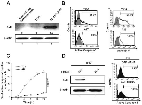
Tumor immune escape is a major obstacle in cancer immunotherapy, but the mechanisms involved remain poorly understood. We have previously developed an immune evasion tumor model using an in vivo immune selection strategy and revealed Akt-mediated immune resistance to antitumor immunity induced by various cancer immunotherapeutic agents. In the current study, we used microarray gene analysis to identify an Akt-activating candidate molecule overexpressed in immune-resistant tumors compared with parental tumors. X-linked lymphocyte-regulated protein pM1 (XLR) gene was the most upregulated in immune-resistant tumors compared with parental tumor cells. Furthermore, the retroviral transduction of XLR in parental tumor cells led to activation of Akt, resulting in upregulation of antiapoptotic proteins and the induction of immune resistance phenotype in parental tumor cells. In addition, we found that transduction of parental tumor cells with other homologous genes from the mouse XLR family, such as synaptonemal complex protein 3 (SCP3) and XLR-related, meiosis-regulated protein (XMR) and its human counterpart of SCP3 (hSCP3), also led to activation of Akt, resulting in the upregulation of antiapoptotic proteins and induction of immune resistance phenotype. Importantly, characterization of a panel of human cervical cancers revealed relatively higher expression levels of hSCP3 in human cervical cancer tissue compared with normal cervical tissue. Thus, our data indicate that ectopic expression of XLR and its homologues in tumor cells represents a potentially important mechanism for tumor immune evasion and serves as a promising molecular target for cancer immunotherapy.


Figures

Figure 1
Figure 1. Characterization of the expression and anti-apoptotic function of XLR in TC-1 P0 and A17 tumor cells
A. Western blot analysis characterizing the XLR protein expression in TC-1 and A17 cells. XLR-specific antibody was used and incubated with goat anti-mouse IgG conjugated to horseradish peroxidase for visualization of XLR protein using Hyperfilm-enhanced chemiluminescence. β-actin was used as a loading control. B. Flow cytometry analysis of apoptotic cell death induced by E7-specific CD8+ T cells at 1:1 ratio for 4 hours. Apoptosis was measured by quantification of active caspase-3 expression and annexin-V staining. C. Kinetics of caspase-3 positive apoptosis after CTL assay described in Fig 1B. D. Western blot analysis to characterize the XLR expression in GFP or XLR siRNA transfected A17 cells and flow cytometry analysis of caspase-3 positive apoptosis induced by E7-specific CD8+ T cells in A17 cells transfected with GFP or XLR siRNA. A17 cells were transfected with 0.5uM GFP or XLR siRNA and then cultured for 48 hours and Western blot analysis was performed. Data shown are representative of three independent experiments.
Figure 2
Figure 2. Functional characterization of TC-1/XLR tumor cell line
A. Western blot analysis to characterize the expression of XLR and E7 in TC-1/no insert and TC-1/XLR cells. B. Flow cytometry analysis to characterize MHC class I expression on TC-1/no insert and TC-1/XLR cells. PE-conjugated anti-mouse H-2Db monoclonal antibody was used to detect MHC class I expression. The isotype antibody was used as the negative control (black profile). C. Intracellular cytokine staining and flow cytometry analysis to determine the number of IFN-γ secreting E7-specific CD8+ T cells induced by TC-1/no insert and TC-1/XLR cells. TC-1/no insert and TC-1/XLR cells were incubated with E7-specific CD8+ T cells at different E:T rations (0.01:1, 0.1:1, and 1:1) for 16 hours. After incubation, cells were stained for CD8 and IFN-γ, and analyzed by flow cytometry analysis. D. Graphical representation of the tumor volume in mice challenged with TC-1/No insert and TC-1/XLR cells with or without vaccination with Vac-Sig/E7/LAMP-1. Data shown are representative of three independent experiments.
Figure 3
Figure 3. Characterization of the signal pathways that may play a role in increased immune resistance in TC-1/XLR tumor cells
A. Western blot analysis to characterize the expression of total Akt, Ser 473 pAkt, total Erk, Thr 202/Tyr 204 pErk, total p38 MAP kinase and Thr 180/Tyr 182 pp38 MAP kinase in the TC-1/no insert and TC-1/XLR cells. B. Western blot analysis to characterize the expression of total Akt, Ser 473 pAkt, total Erk, Thr 202/Tyr 204 pErk, total p38 MAP kinase and Thr 180/Tyr 182 pp38 MAP kinase in TC-1/XLR cells treated with the various inhibitors. TC-1/XLR cells were incubated with DMSO, PI3K inhibitor LY294002, p38 MAP kinase inhibitor SB203580 or Erk inhibitor PD98059 for 18 hours prior to lysate preparation. C. Representative flow cytometry data for detection of apoptotic TC-1/XLR cells in the presence or absence of E7-specific CD8+ T cells after treatment of DMSO or each inhibitor. TC-1/XLR cells were pretreated with the various inhibitors and incubated with E7-specific CD8+ T cells at 1:1 ratio. After 4 hour incubation, apoptosis was evaluated The data are representative of three separate experiments. D. Bar graph depicting the percentage of apoptotic TC-1/XLR cells.
Figure 4
Figure 4. Characterization of anti-tumor effects generated by E7 specific CD8+ T cell adoptive transfer combined with AKT inhibitor API-2 treatment
A. Western blot analysis to characterize the expression of p-AKT, AKT, Bcl-XL and Bcl-2 in DMSO or Akt inhibitor API-2 treated TC-1/XLR cells. B. Bar graph representing tumor volumes from tumor challenged mice treated with or without API-2 in the presence or absence of E7-specific CD8+ T cells on 10 days after tumor challenge. Tumor volumes from TC-1/XLR tumor were recorded twice per week for 10 days following adoptive transfer.
Figure 5
Figure 5. Characterization of expression of XLR family genes in the TC-1/No insert and TC-1/XLR tumor cells
A. Western blot analysis to characterize the expression of SCP3 in TC-1/no insert and TC-1/SCP3 and HA expression in the TC-1/no insert and TC-1/XMR/HA cells and p-AKT, total Akt, Mcl-1, Bcl-xL and Bcl-2 expression in TC-1 cells expressing the XLR homologs. B. Flow cytometry analysis to characterize MHC class I expression on TC-1 cells expressing the XLR homologs compared to TC-1/no insert cells and in vitro T cell activation to determine the number of IFN-γ secreting E7-specific CD8+ T cells induced by TC-1 cells expressing the XLR homologs compared to TC-1/no insert cells. MFI; mean fluorescence intensity. C. Bar graph depicting percentage of apoptotic TC-1 cells expressing the XLR homologs compared to TC-1/no insert cells incubated with E7-specific CD8+ T cells at 1:1 ratio for 4 hours. D. Graphical representation of the tumor volume in mice challenged with TC-1/XLR, TC-1/SCP3 or TC-1/XMRtumor cells with or without adoptive transfer of E7-specific CD8+ T cells. Data shown are representative of three independent experiments.
Figure 6
Figure 6. Characterization of the apoptotic cell death of 293Db/no insert and 293Db/SCP3 induced by E7 CTL killing in vitro
A. Western blot analysis to characterize the expression of SCP3 in 293Db/no insert and 293Db/hSCP3 cells. B. Western blot analysis to characterize the expression of p-AKT, Bcl-2 and Bcl-xL in 293Db/no insert and 293Db/hSCP3 cells. β-actin was used as a loading control. C. Representative flow cytometry data demonstrating the percentage of apoptotic 293Db/no insert and 293Db/hSCP3 cells. 293Db/no insert and 293Db/hSCP3 cells were incubated with E7-specific CD8+ T cells at different E:T ratios (1:1 or 5:1) for 4 hours. After incubation, the percentage of apoptotic cells was analyzed using intracellular flow cytometry analysis for active caspase-3 expression. D. Graphical representation of the percentage of apoptotic cells among 293Db/no insert and 293Db/hSCP3 cells at different E:T ratios.

References

    1. Rosenberg SA, Yang JC, Restifo NP. Cancer immunotherapy: moving beyond current vaccines. Nat Med. 2004;10:909–15. - PMC - PubMed
    1. Lin KY, Guarnieri FG, Staveley-O’Carroll KF, et al. Treatment of established tumors with a novel vaccine that enhances major histocompatibility class II presentation of tumor antigen. Cancer Res. 1996;56:21–6. - PubMed
    1. Wu TC. The role of vascular cell adhesion molecule-1 in tumor immune evasion. Cancer Res. 2007;67:6003–6. - PMC - PubMed
    1. Wu TC, Guarnieri FG, Staveley-O’Carroll KF, et al. Engineering an intracellular pathway for major histocompatibility complex class II presentation of antigens. Proc Natl Acad Sci U S A. 1995;92:11671–5. - PMC - PubMed
    1. Noh KH, Kang TH, Kim JH, et al. Activation of Akt as a Mechanism for Tumor Immune Evasion. Mol Ther. 2008;17:439–47. - PMC - PubMed

Publication types