. 2010 Jun 18;285(25):19173-83.
doi: 10.1074/jbc.M110.102582. Epub 2010 Apr 15.

A novel native store-operated calcium channel encoded by Orai3: selective requirement of Orai3 versus Orai1 in estrogen receptor-positive versus estrogen receptor-negative breast cancer cells

Affiliations

A novel native store-operated calcium channel encoded by Orai3: selective requirement of Orai3 versus Orai1 in estrogen receptor-positive versus estrogen receptor-negative breast cancer cells

Rajender K Motiani et al. J Biol Chem. .

Abstract

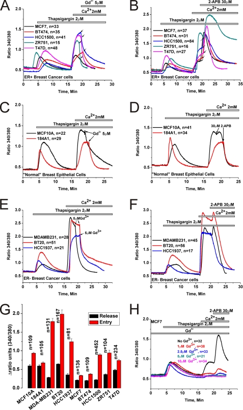
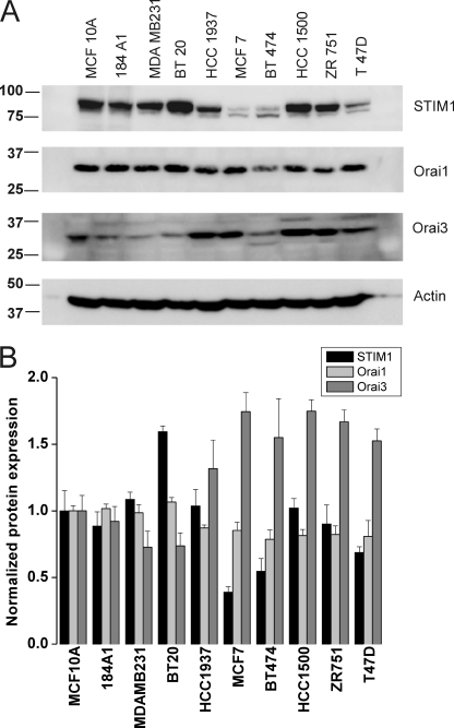
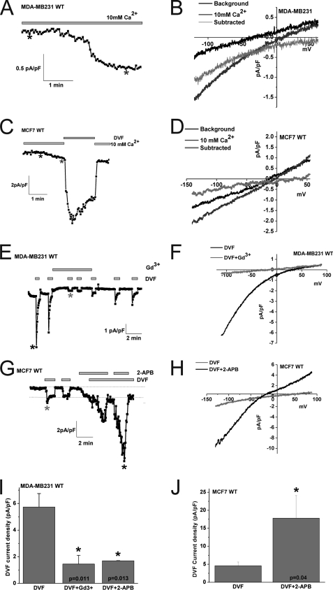
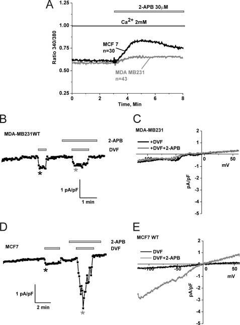
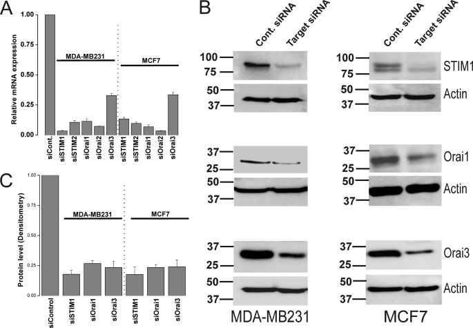
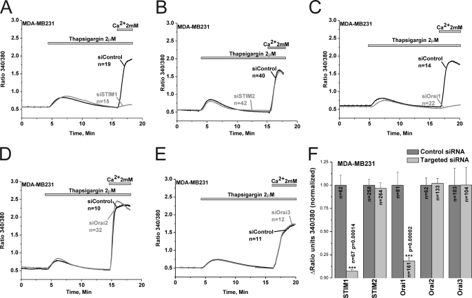
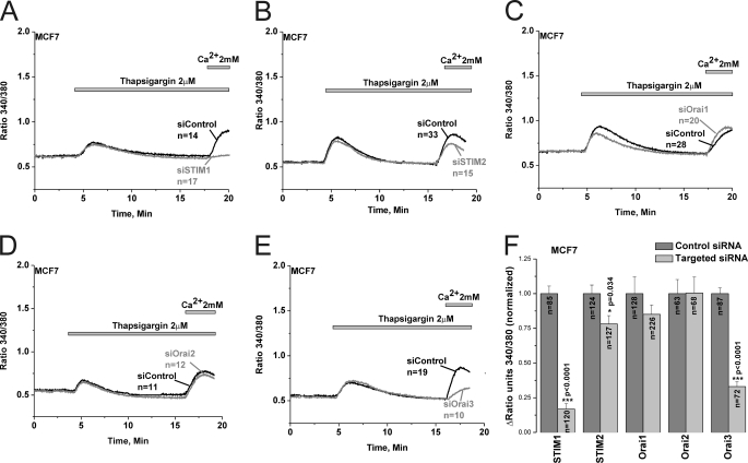
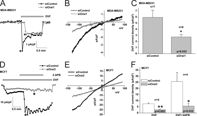
Store-operated calcium (Ca(2+)) entry (SOCE) mediated by STIM/Orai proteins is a ubiquitous pathway that controls many important cell functions including proliferation and migration. STIM proteins are Ca(2+) sensors in the endoplasmic reticulum and Orai proteins are channels expressed at the plasma membrane. The fall in endoplasmic reticulum Ca(2+) causes translocation of STIM1 to subplasmalemmal puncta where they activate Orai1 channels that mediate the highly Ca(2+)-selective Ca(2+) release-activated Ca(2+) current (I(CRAC)). Whereas Orai1 has been clearly shown to encode SOCE channels in many cell types, the role of Orai2 and Orai3 in native SOCE pathways remains elusive. Here we analyzed SOCE in ten breast cell lines picked in an unbiased way. We used a combination of Ca(2+) imaging, pharmacology, patch clamp electrophysiology, and molecular knockdown to show that native SOCE and I(CRAC) in estrogen receptor-positive (ER(+)) breast cancer cell lines are mediated by STIM1/2 and Orai3 while estrogen receptor-negative (ER(-)) breast cancer cells use the canonical STIM1/Orai1 pathway. The ER(+) breast cancer cells represent the first example where the native SOCE pathway and I(CRAC) are mediated by Orai3. Future studies implicating Orai3 in ER(+) breast cancer progression might establish Orai3 as a selective target in therapy of ER(+) breast tumors.


Figures

FIGURE 1.
FIGURE 1.
Pharmacology of SOCE in ER+ and ER breast cell lines. Cells were stimulated by 2 μm thapsigargin in the absence of extracellular Ca2+ to passively deplete the intracellular Ca2+ stores followed by restoration of Ca2+ (2 mm) to the extracellular space to assess SOCE. The effects of subsequent addition of either 5 μm Gd3+ or 30 μm 2-APB on thapsigargin-activated SOCE in ER+ breast cancer cell lines (A and B), ER normal breast epithelial cells (C and D) and ER breast cancer cell lines (E and F) are shown. Arrows indicate for every cell line the exact moment of Gd3+ and 2-APB addition. G, the extent of SOCE as well as Ca2+ release from internal stores in response to thapsigargin was calculated in a large number of cells, for every cell line, by subtracting the basal Fura2 ratio value from that obtained after achieving either maximal peak of release in nominally free Ca2+ solution or maximal Ca2+ entry upon Ca2+ restoration to the external space; mean of data ± S.E. are shown. H, MCF7 cells were incubated in the presence of varying concentrations of Gd3+ (1–10 μm) to block SOCE followed by 30 μm 2-APB to determine the extent of potentiation; a control condition without Gd3+ is also included. Data are representative of at least four independent experiments.
FIGURE 2.
FIGURE 2.
Western blot analysis of STIM1, Orai1, and Orai3 expression in breast cancer cells. A, all 10 breast cancer cells lines were lysed, and 50 μg of proteins each were loaded in the same gels. After transfer, membranes were probed with specific antibodies against either STIM1, Orai1, or Orai3 followed by the appropriate secondary antibody coupled to peroxidase as described under “Materials and Methods,” and proteins bands were visualized using the ECL kit. B, blots obtained from 3–4 independent experiments were analyzed using Image J software and densitometric ratios to corresponding actin were calculated. All densitometric values were then normalized to those of the normal cell line MCF10A.
FIGURE 3.
FIGURE 3.
Whole-cell SOCE currents in MDA-MB231 ER and MCF7 ER+ breast cancer cells. Wild-type MDA-MB231 cells (A) or wild-type MCF7 cells (C) were dialyzed with a pipette solution containing 12 mm BAPTA to induce store depletion and whole-cell currents were measured in the presence of 10 mm extracellular Ca2+. A small inwardly rectifying ICRAC-like current developed in both cell types. B and D show the current/voltage (I/V) relationships of these Ca2+ currents in MDA-MB231 and MCF7 cells, respectively; the sweeps after break-in (black) and those when the current has developed (dark gray) were taken where shown by asterisks. The light gray sweep represents the subtraction of the “black” sweep from the “dark gray” sweep. For MCF7 (C), the trace depicted also shows amplification of the current by switching to a divalent-free (DVF) solution. Monovalent ICRAC measured in MDA-MB231 cells (E) and MCF7 cells (G) under DVF conditions is shown. Monovalent ICRAC currents measured from MDA-MB231 show substantial “depotentiation” in DVF solutions while those measured in MCF7 have a less pronounced depotentiation. Monovalent ICRAC in MCF7 is potentiated by 2-APB and this is accompanied by a change in the I/V relationship of the current (left shift of reversal potential and outward component. H, in this case, background currents were not subtracted). The I/V curve of monovalent ICRAC in MDA-MB231 is shown in F. Monovalent ICRAC in MDA-MB231 is inhibited by Gd3+ (5 μm) and 2-APB (50 μm), and data are summarized in I (n = 5). J, data summary of monovalent ICRAC current density in MCF7 cells before and after 2-APB (50 μm) potentiation (n = 7).
FIGURE 4.
FIGURE 4.
2-APB activates Ca2+ entry and membrane currents in MCF7 cells in the absence of store depletion. A, MDA-MB231 and MCF7 cells were loaded with Fura2 and incubated in an HBSS solution containing 2 mm Ca2+ followed by stimulation with 30 μm 2-APB; only MCF7 show a significant Ca2+ entry upon 2-APB stimulation (similar results were obtained with 50 μm 2-APB). Using a pipette solution where Ca2+ was buffered to the physiological concentration of 100 nm, whole cell recording in MDA-MB231 showed no increase in DVF currents after addition of 50 μm 2-APB (B and C) while MCF7 showed substantial current activation in response to the same concentration of 2-APB (D and E). The I/V curves represented in C and E were taken where indicated by the color-coded asterisks in B and D, respectively.
FIGURE 5.
FIGURE 5.
Protein and mRNA knockdown of STIM and Orai isoforms in MDA-MB231 and MCF7 cells. MDA-MB231 and MCF7 cells were transfected with either control non-targeting siRNA or specific siRNA against STIM1, STIM2, Orai1, Orai2, or Orai3 and mRNA were extracted 72-h post-transfection, reverse transcribed, and assayed using real-time PCR as described under “Materials and Methods.” Each specific siRNA caused a significant decrease of its corresponding target mRNA (A). Data represent average ± S.E. from three independent transfections analyzed in duplicates. Western blotting analysis using specific antibodies was conducted to assess the protein knockdown of STIM1, Orai1, and Orai3 after transfection with specific siRNA and non-targeted control siRNA in MDA-MB231 and MCF7 cells; the corresponding anti-actin loading controls are also shown (B). Data are representative of three independent transfections and statistical analysis of protein knockdown is included in the text and depicted in C.
FIGURE 6.
FIGURE 6.
SOCE in MDA-MB231 cells is mediated by STIM1 and Orai1. Thapsigargin-activated SOCE in MDA-MB231 cells was measured using Fura2 and the standard “Ca2+ off/Ca2+ on” protocol in cells transfected with either specific siRNA against STIM1 (A), STIM2 (B), Orai1 (C), Orai2 (D), or Orai3 (E) or control non-targeted siRNA performed on the same day; measurements were performed on day 3 post-transfection. Data are representative of 6–15 independent experiments from 2–4 independent transfections. The effect of siRNA against STIM and Orai isoforms and corresponding control non-targeted siRNA on SOCE in MDA-MB231 was statistically analyzed on cells from different independent experiments and depicted in F.
FIGURE 7.
FIGURE 7.
SOCE in MCF7 cells is mediated by STIM1/2 and Orai3. Thapsigargin-activated SOCE in MCF7 cells was measured using Fura2 and the standard “Ca2+ off/Ca2+ on” protocol in cells transfected with either specific siRNA against STIM1 (A), STIM2 (B), Orai1 (C), Orai2 (D), or Orai3 (E) or control non-targeted siRNA performed on the same day; measurements were performed on day 3 post-transfection. The effect of siRNA against STIM and Orai isoforms and corresponding control non-targeted siRNA on SOCE in MCF7 was statistically analyzed on cells from different independent experiments and depicted in F.
FIGURE 8.
FIGURE 8.
Orai1 and Orai3 mediate ICRAC in MDA-MB231 and MCF7 cells, respectively. A, Orai1 knockdown causes inhibition of monovalent ICRAC measured using DVF pulses as compared with non-targeted control siRNA conditions. The I/V relationships of control siRNA and Orai1-targeted siRNA conditions in MDA-MB231 cells are shown in B, and statistical analysis on the effect of Orai1 knockdown on monovalent ICRAC (n = 7) is represented in C. D, Orai3 knockdown causes inhibition of monovalent ICRAC in MCF7 cells as well as an inhibition of 2-APB-mediated potentiation of this current. The I/V relationships of control siRNA and Orai3-targeted siRNA conditions in MCF7 cells after 2-APB addition are shown in E. Statistical analysis on the effect of Orai3 knockdown on monovalent ICRAC in MCF7 cells (n = 4) before and after 2-APB addition is represented in F. Throughout, background currents were not subtracted.

References

    1. Glass A. G., Lacey J. V., Jr., Carreon J. D., Hoover R. N. (2007) J. Natl. Cancer Inst. 99, 1152–1161 - PubMed
    1. Jemal A., Ward E., Thun M. J. (2007) Breast Cancer Res. 9, R28. - PMC - PubMed
    1. Bai Z., Gust R. (2009) Archiv der Pharmazie 342, 133–149 - PubMed
    1. Hurvitz S. A., Finn R. S. (2009) Future Oncology 5, 1015–1025 - PubMed
    1. Parekh A. B., Putney J. W., Jr. (2005) Physiol. Rev. 85, 757–810 - PubMed

Publication types