Review
. 2010 May 1;51 Suppl 1(Suppl 1):51S-65S.
doi: 10.2967/jnumed.109.068163. Epub 2010 Apr 15.

Imaging atherosclerosis and vulnerable plaque

Affiliations
Review

Imaging atherosclerosis and vulnerable plaque

Mehran M Sadeghi et al. J Nucl Med. .

Abstract

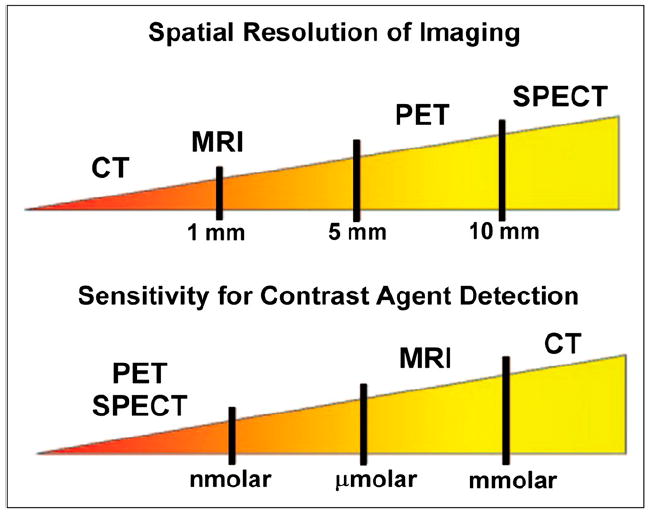
Identifying patients at high risk for an acute cardiovascular event such as myocardial infarction or stroke and assessing the total atherosclerotic burden are clinically important. Currently available imaging modalities can delineate vascular wall anatomy and, with novel probes, target biologic processes important in plaque evolution and plaque stability. Expansion of the vessel wall involving remodeling of the extracellular matrix can be imaged, as can angiogenesis of the vasa vasorum, plaque inflammation, and fibrin deposits on early nonocclusive vascular thrombosis. Several imaging platforms are available for targeted vascular imaging to acquire information on both anatomy and pathobiology in the same imaging session using either hybrid technology (nuclear combined with CT) or MRI combined with novel probes targeting processes identified by molecular biology to be of importance. This article will discuss the current state of the art of these modalities and challenges to clinical translation.


Figures

FIGURE 1
FIGURE 1
RP782 imaging of MMP activation in vascular remodeling. RP782 micro-SPECT (A), CTA (B), and fused micro-SPECT/CT (C) images at 3 wk after left carotid injury in apoE−/− mice demonstrate enhanced RP782 uptake in injured left as compared with control right carotid arteries. Quantification of carotid RP782 uptake at different time points after injury is shown in (D). S = sagittal slices; C = coronal slices; T = transverse slices; w = weeks. (Reprinted with permission of (6).)
FIGURE 2
FIGURE 2
Contrast CT (left), micro-SPECT (center), and fused SPECT/CT (right) images of apoE−/− mice fed Western diet for more than 16 wk. Imaging showed no focal aortic arch hot spots in mice injected with a nonspecific IgG antibody (nIgG) probe (top row), whereas all mice injected with targeted LOX-1 probe had hot spots in aortic arch (lower row). Results were confirmed by ex vivo phosphor imaging of excised aortas. Sudan IV staining demonstrated comparable plaques between the 2 groups.
FIGURE 3
FIGURE 3
(A) Six reconstructed slices from in vivo hybrid small-animal SPECT/CT scan after injection of 99mTc annexin AV into 62-wk apoE−/− mouse fed high-fat diet and showing uptake of tracer in aortic arch (red arrows). Image on right shows excised aorta imaged ex vivo. (B) Immunohistochemical stained sections through aorta shows American Heart Association class IV lesion with lipid core, prevalent macrophages, and TUNEL-positive nuclei. (C) Correlations between percentage injected dose (%ID) and both macrophages and TUNEL-positive cells. (Reprinted from (35).)
FIGURE 4
FIGURE 4
(Top left) Reconstructed slices from in vivo hybrid small-animal SPECT/CT scan after injection of 99mTc-labeled MPI into representative mice from 5 groups: control, apoE−/− fed high-fat diet (ChApo∈−/−), apoE−/− fed normal chow, LDLR−/− fed high-fat diet (ChLDLR−/−), and LDLR−/− fed normal diet. Black arrows identify aorta, and red arrows identify tracer uptake in aortic arch (on SPECT and fused SPECT/CT), with greatest amount seen in apoE−/− mouse fed high-fat diet. Scans of control mouse are negative. (Top right) Quantitative histologic analysis of MMP-2, MMP-9, and macrophages (Mac-3) for 4 groups of atherosclerotic mice, and control. (Bottom) Histopathologic and immunohistochemical staining of sections from aortae from 5 groups. (Reprinted from (43).)
FIGURE 5
FIGURE 5
(A) Time-of-flight MR angiogram 30 min after balloon stretch injury shows patent femoral arteries. Left artery was treated with αvβ3-integrin– targeted paramagnetic nanoparticles with rapamycin, and saline was used for right artery. (B and C) MR angiograms 2 wk after injury and treatment, with arrows identifying regions of intraluminal plaque caused by balloon overstretch injury. In B, right artery, which has arterial plaque, was treated with αvβ3-integrin–targeted nanoparticles without drug, and widely patent left artery was treated with αvβ3-integrin– targeted nanoparticles with rapamycin. In C, widely patent right artery was treated with αvβ3-integrin–targeted nanoparticles with rapamycin, and partially occluded left artery was treated with nontargeted nanoparticles with rapamycin. (D and E) Graphs of average (D) and maximum average (E) stenosis within injured and treated femoral arteries of New Zealand White rabbits 2 wk after balloon injury. Arterial segments were flash-frozen in optimal-cutting-temperature compound, and alternate 7-μm sections were used for morphologic analysis (hematoxylin and eosin staining). (F) Area at risk of injured endothelium quantified on vascular en face preparations stained with Carstair stain. Normal, uninjured endothelium is yellow, and injured endothelium with fibrin deposition is red. (G) Quantitation of injured endothelium in area at risk (100% = 1-cm excised vessel segment). Digitized images were analyzed on areas that had undergone balloon overstretch injury and were treated with αvβ3-integrin–targeted nanoparticles with 0.4 mol% rapamycin (n = 12) or saline control (n = 12). Vessels were excised on postinterventional days 1, 7, 14, and 28 (n = 3 per group and time point). (Adapted with permission of (91).)
FIGURE 6
FIGURE 6
(A) MRI of abdominal aorta shows false-colored overlay of percentage signal enhancement at time of treatment (left) and 1 wk after treatment (right). (B) Platelet endothelial cell adhesion molecule (PECAM)–stained section (×4) of abdominal aorta from hyperlipidemic rabbit shows adventitia, media, and plaque. Higher-magnification inset (×20) shows that microvessels were predominantly in adventitia associated with thickening neointima. Neovessels were generally not in regions where plaque progression was minimal or nonexistent in this cohort of rabbits. Arrowheads illustrate type of PECAM microvessels counted within each section to assess fumagillin antiangiogenic effects. Larger, mature vessels positively staining for PECAM were not included in these estimates. (C) Graph of aortic MRI signal enhancement averaged over all imaged slices at time of treatment (black bars) and 1 wk after treatment (white bars). Solid lines indicate individual animal’s response to treatment over 7-d period. (D) Graph showing that number of neovascular vessels within adventitia was reduced (*P < 0.06; P < 0.05) in fumagillin-treated rabbits over proximal half of aorta (i.e., renal artery to diaphragm), which correlated with region of greatest MRI signal and fumagillin response in imaging studies. (Adapted with permission of (82).)
FIGURE 7
FIGURE 7
Illustration of relative spatial resolution of common imaging techniques, along with their sensitivities.

References

    1. Pasterkamp G, Fitzgerald PF, de Kleijn DP. Atherosclerotic expansive remodeled plaques: a wolf in sheep’s clothing. J Vasc Res. 2002;39:514–523. - PubMed
    1. Johnson LL, Schofield LM, Verdesca SA, et al. In vivo uptake of radiolabeled antibody to proliferating smooth muscle cells in a swine model of coronary stent restenosis. J Nucl Med. 2000;41:1535–1540. - PubMed
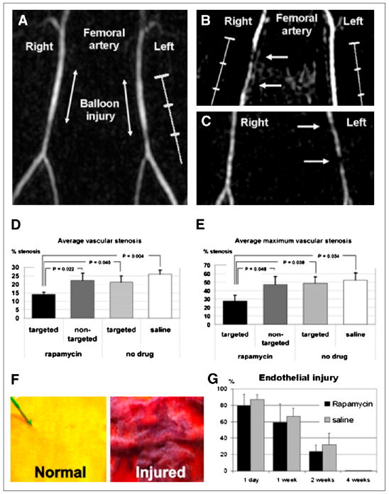
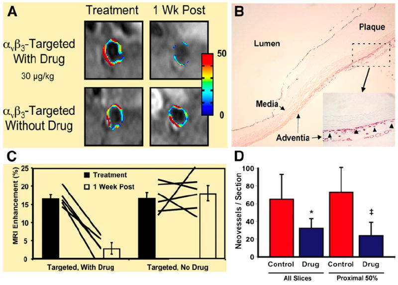
    1. Sadeghi MM, Krassilnikova S, Zhang J, et al. Detection of injury-induced vascular remodeling by targeting activated alphavbeta3 integrin in vivo. Circulation. 2004;110:84–90. - PubMed
    1. Zhang J, Krassilnikova S, Gharaei AA, et al. Alphavbeta3-targeted detection of arteriopathy in transplanted human coronary arteries: an autoradiographic study. FASEB J. 2005;19:1857–1859. - PubMed
    1. Schafers M, Riemann B, Kopka K, et al. Scintigraphic imaging of matrix metalloproteinase activity in the arterial wall in vivo. Circulation. 2004;109:2554–2559. - PubMed

MeSH terms