

CT features of parotid gland oncocytomas: a study of 10 cases and literature review
- PMID: 20395389
- PMCID: PMC7966095
- DOI: 10.3174/ajnr.A2090
CT features of parotid gland oncocytomas: a study of 10 cases and literature review
Abstract
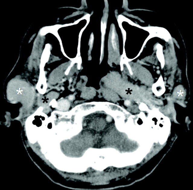
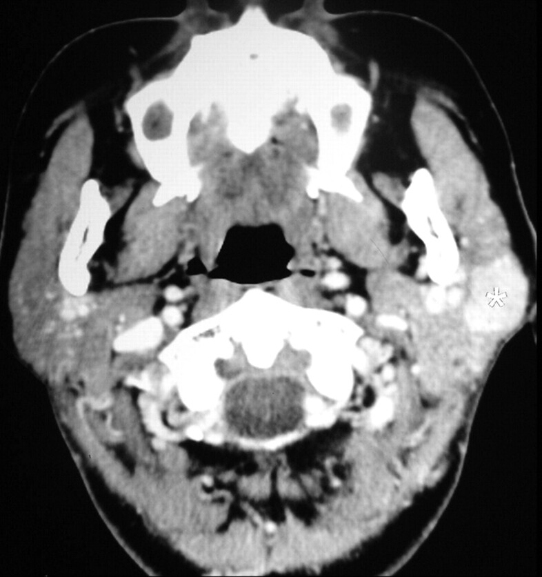
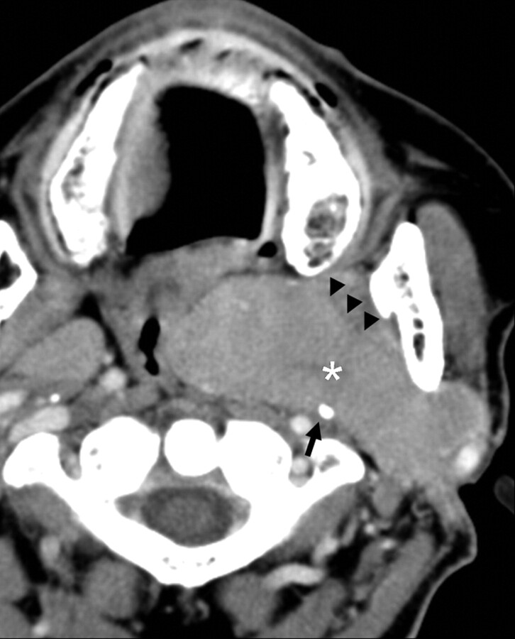
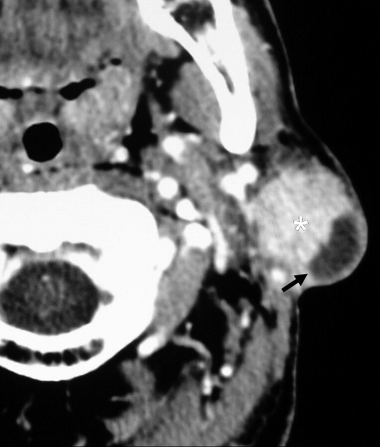
Oncocytomas of the salivary glands are rare benign epithelial tumors which occur most commonly in the parotid gland. The aim of our study was to characterize the clinical-radiologic-pathologic spectrum of parotid oncocytomas in a series of 10 cases seen in our institution between January 2003 and November 2008. The CT features of parotid oncocytomas in the largest imaging series of this rare but important benign lesion include a well-defined enhancing tumor with a "deformable" appearance when large, and a non-enhancing curvilinear cleft or cystic component. These CT findings are potentially helpful in distinguishing these benign lesions from other parotid tumors in clinical scenarios that preclude surgical resection or when biopsy results are non-diagnostic. Further studies are advocated to validate the specificity and positive predictive value of these imaging features.
Figures

References
-
- Hamperl H. Benign and malignant oncocytoma. Cancer 1962;15:1019–27 - PubMed
-
- Lee YY, Wong KT, King AD, et al. . Imaging of salivary gland tumors. Eur J Radiol 2008;66:419–36 - PubMed
-
- Barnes L, Eveson JW, Reichart P, et al., eds. World Health Organization Classification of Tumors: Pathology and Genetics of Head and Neck Tumors. Lyon, France: International Agency for Research on Cancer Press; 2005:237,268
-
- Capone RB, Ha PK, Westra WH, et al. . Oncocytic neoplasm of the parotid gland: a 16-year institutional review. Otolaryngol Head Neck Surg 2002;126:657–62 - PubMed
-
- Politi M, Toro C, Cian R, et al. . Multifocal adenomatous oncocytic hyperplasia of the parotid gland. Oral Oncol Extra 2005;41:183–87
Publication types
MeSH terms
LinkOut - more resources
Full Text Sources
Medical