. 2010 Aug 12;116(6):915-25.
doi: 10.1182/blood-2009-11-254334. Epub 2010 Apr 15.

Comprehensive analysis of lymph node stroma-expressed Ig superfamily members reveals redundant and nonredundant roles for ICAM-1, ICAM-2, and VCAM-1 in lymphocyte homing

Affiliations

Comprehensive analysis of lymph node stroma-expressed Ig superfamily members reveals redundant and nonredundant roles for ICAM-1, ICAM-2, and VCAM-1 in lymphocyte homing

Rémy T Boscacci et al. Blood. .

Abstract

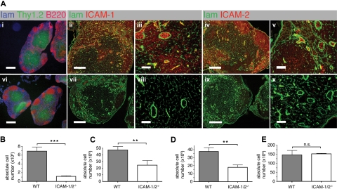
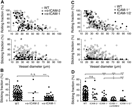
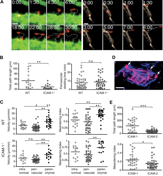
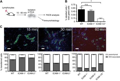
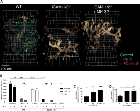
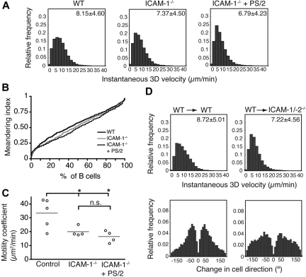
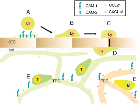
Although it is well established that stromal intercellular adhesion molecule-1 (ICAM-1), ICAM-2, and vascular cell adhesion molecule-1 (VCAM-1) mediate lymphocyte recruitment into peripheral lymph nodes (PLNs), their precise contributions to the individual steps of the lymphocyte homing cascade are not known. Here, we provide in vivo evidence for a selective function for ICAM-1 > ICAM-2 > VCAM-1 in lymphocyte arrest within noninflamed PLN microvessels. Blocking all 3 CAMs completely inhibited lymphocyte adhesion within PLN high endothelial venules (HEVs). Post-arrest extravasation of T cells was a 3-step process, with optional ICAM-1-dependent intraluminal crawling followed by rapid ICAM-1- or ICAM-2-independent diapedesis and perivascular trapping. Parenchymal motility of lymphocytes was modestly reduced in the absence of ICAM-1, while ICAM-2 and alpha4-integrin ligands were not required for B-cell motility within follicles. Our findings highlight nonredundant functions for stromal Ig family CAMs in shear-resistant lymphocyte adhesion in steady-state HEVs, a unique role for ICAM-1 in intraluminal lymphocyte crawling but redundant roles for ICAM-1 and ICAM-2 in lymphocyte diapedesis and interstitial motility.


Figures

Figure 1
Figure 1
PLN architecture and cellularity of lymphoid organs in WT and ICAM-1/2−/− mice. (A) Immunofluorescent staining of PLN sections of WT (i-v) and ICAM-1/2−/− (vi-x) mice. i,vi: laminin (lam; blue), Thy1.2 (green), and B220 (red). Scale bar = 400 μm. ii,iii,vii,viii: laminin (lam; green) and ICAM-1 (red). Scale bar = 200 μm (ii,vii) or 40 μm (iii,viii). iv,v,ix,x: laminin (lam; green) and ICAM-2 (red). Scale bar = 200 μm (iv,ix) or 40 μm (v,x). (B-E) Absolute cell numbers per cervical, brachial, axillary, inguinal, paraaortal, and popliteal PLNs (B), MLN (C), PP (D), and spleen (E) of WT and ICAM-1/2−/− mice, represented as mean plus or minus SEM pooled from 3 (PLN, MLN, spleen) or 4 (PP) independent experiments. n.s. indicates not significant. **P < .01; ***P < .0001.
Figure 2
Figure 2
IVM analysis of lymphocyte rolling and shear-resistant firm adhesion in mAb-blocked WT and ICAM-1−/−, ICAM2−/− and ICAM-1/2−/− PLN microvasculature. (A) Rolling (top panel) and sticking fractions (bottom panel) of lymphocytes in WT PLN microvessels as a function of vessel diameter and in the presence of blocking antibodies against ICAM-1 or ICAM-2. Each dot represents 1 venule. Pooled from 3 (+anti–ICAM-2), 18 (+anti–ICAM-1), and 27 (no Abs) WT animals per condition with 5 (+anti–ICAM-2), 59 (+anti–ICAM-1) and 126 (no Abs) venules analyzed. (B) Sticking fractions of lymphocytes in WT PLN microvessels as in panel A. Each dot represents 1 venule. Bars represent mean values. n.s. indicates not significant. ***P < .0001. (C) Rolling (top panel) and sticking fractions (bottom panel) of lymphocytes in WT, ICAM-1−/−, and ICAM-1/2−/− PLN microvessels as a function of vessel diameter. Each dot represents 1 venule. Pooled from 6 ICAM-1−/−, 6 ICAM-1/2−/−, and 27 WT animals with 45 (ICAM-1−/−), 42 (ICAM-1/2−/−), and 126 (WT) venules analyzed. (D) Sticking fractions of lymphocytes in WT, ICAM-2−/− (4 animals, 13 venules analyzed), ICAM-1−/−, ICAM-1−/− + anti–ICAM-2 (4 animals, 29 venules analyzed) and ICAM-1/2−/− PLN microvessels as in panel C. Each dot represents 1 venule. Numbers of WT, ICAM-1−/−, and ICAM-1/2−/− animals and analyzed venules as in panel C. Bars represent mean values. n.s. indicates not significant. *P < .05; ***P < .0001.
Figure 3
Figure 3
A nonredundant role for ICAM-1 for intraluminal T-cell crawling, but not transendothelial migration or perivascular trapping. (A) Representative micrographs of directly egressing (left panels) or crawling (right panels) T cells transmigrating through HEVs. Time in minutes and seconds; scale bar = 10 μm. (B left panel) Total path length of crawling T cells in WT or ICAM-1−/− PLNs. (Right panel) Perivascular dwelling of transmigrated T cells in WT or ICAM-1−/− PLNs. (C top panels) Mean track velocity (right) and meandering index (MI; left) of crawling intraluminal T cells, as well as perivascular and parenchymal T cells in WT PLNs. The low velocity and MI are indicative of the perivascular slowing down of transmigrated T cells. (Bottom panels) Mean track velocity (right) and MI (left) of intraluminal perivascular and parenchymal T cells in ICAM-1−/− PLNs. (D) Immunofluorescent image of transmigrated T cells (red, arrows) around a MECA-79+ HEV (pink) 15 minutes after transfer. Transferred cells are in close proximity to the laminin+ basement membrane (blue). Scale bar = 10 μm. (E) Total path length (top panel) and MI (bottom panel) of naive T cells migrating on 2D surfaces coated with CCL21 + ICAM-1/Fc or ICAM-2/Fc. Tracks were recorded for 20 minutes. One experiment of 3 is shown. n.s. indicates not significant. *P < .05; **P < .001; ***P < .0001.
Figure 4
Figure 4
Reduced homing in the absence of ICAM-1 and 2. (A) Schematic outline of short-term homing experiment. Fluorescently labeled lymphocytes were injected and allowed to home for 15, 30, or 60 minutes. PLNs were analyzed by flow cytometry or immunohistology. (B) Flow cytometric analysis of homing efficiency in the absence of ICAM-1 or ICAM-1 and ICAM-2. PLNs were analyzed 60 minutes after cell transfer. The percentage of fluorescently labeled lymphocytes of the total PLN cell population is depicted. Pooled from 4 independent experiments. (C top panel) Immunofluorescent analysis of PLN sections at 15, 30, and 60 minutes after cell transfer. Transferred lymphocytes are labeled in red, HEVs (MECA-79+) in green, and ERTR7 (in the PLN parenchyma and the HEV basement membrane) in blue. Cells within the ERTR7+ ring around HEVs were defined as vessel associated and cells outside as parenchymal. Scale bar = 100 μm. (Bottom panel) Localization of transferred lymphocytes in WT, ICAM-1−/−, ICAM-2−/−, and ICAM-1/-2−/− PLNs at 15, 30, and 60 minutes after cell transfer. Absolute numbers per section were normalized to 100%. Bars represent mean plus or minus SEM pooled from 3 to 5 independent experiments.
Figure 5
Figure 5
3-dimensional quantitative immunofluorescence (3DQIF) of lymphocyte homing in the absence of ICAM-1, ICAM-2 and VCAM-1 during lymphocyte trafficking. (A) Representative 3D reconstructions of WT, ICAM-1/2−/−, and ICAM-1/2−/− + anti–VCAM-1 mAb (MK2.7) PLNs after adoptive transfer of untreated lymphocytes (green), or treated with blocking anti–α4-integrin (PS/2, blue) or anti–LFA-1 (FD441.8, red). The MECA79+ HEV network is shown in brown. One square line corresponds to 50 μm. (B) Absolute cell counts per mm3 in control and ICAM-1/2−/− PLNs in the presence of blocking mAbs. In some experiments, WT or ICAM-1/2−/− mice were pretreated with anti–VCAM-1 (MK2.7). Pooled from 5 experiments with 15 (ICAM-1/2−/−) and 16 (WT) independent scans. (C) Intravascular frequency of adoptively transferred untreated lymphocytes in WT and ICAM-1/2−/− PLNs (right bar), and FD441.8-treated lymphocytes in WT PLNs (middle bar). (D) Perivascular frequency of adoptively transferred untreated lymphocytes in WT and ICAM-1/2−/− PLNs (right bar), and FD441.8-treated lymphocytes in WT PLNs (middle bar). Total cell counts in panels C and D were normalized to 100%. Data in panels C and D are pooled from 5 independent experiments. Bars represent mean plus minus SEM. n.s. indicates not significant. *P < .05; **P < .001; ***P < .0001.
Figure 6
Figure 6
Follicular B-cell migration in absence of ICAM-1. (A) Instantaneous 3D velocity of follicular B cells in WT and ICAM-1−/− PLNs, treated or not with the anti-α4 blocking mAb PS/2. The mean velocity plus or minus SEM is indicated in the top right corner in μm/min. B cells are significantly slower in absence of ICAM-1 (P < .001), and their velocity can be further decreased after treatment with PS/2 (P < .001). (B) Normalized, cumulative meandering indices of B cells migrating in WT and ICAM-1−/− PLNs, treated or not with the anti-α4 blocking mAb PS/2. (C) Motility coefficients of B cells migrating in WT and ICAM-1−/− PLNs, treated or not with the anti-α4 blocking mAb PS/2. Absence of ICAM-1 significantly reduces the motility coefficient. n.s. indicates not significant. *P < .05. Data are pooled from 2 to 4 independent experiments yielding 4 to 5 image sequences containing 653, 345, and 340 cell tracks for WT, ICAM-1−/−, and ICAM-1−/− + PS/2 PLNs, respectively. (D) Instantaneous 3D velocity (top panels) and change in cell direction distribution (turning angles, bottom panels) of follicular B cells in PLNs of WT and ICAM-1/-2−/− BM chimeras. For WT→WT BM chimera, 1 mouse per 4 image sequences per 442 cell tracks was analyzed, while for WT→ICAM-1-2−/− BM chimera, we examined 2 mice per 4 image sequences per 389 tracks.
Figure 7
Figure 7
Proposed role for ICAM-1 and ICAM-2 during lymphocyte trafficking to and within PLNs. Blood-borne lymphocytes undergo rapid firm arrest in a largely ICAM-1 and ICAM-2-dependent manner, with a residual contribution of VCAM-1, in particular in the smallest HEVs (A). Approximately one-third of adherent T cells subsequently crawl on the luminal HEV surface, independent of the direction of the blood flow, in an ICAM-1-dependent manner (B). The rapid transmigration event (∼ 1.5 minutes) can occur in the absence of endothelial ICAM-1 and ICAM-2 (C), and is followed in the majority of cells by “perivascular trapping” of the cells, perhaps because of entanglement with the basement membrane underlying the HEV and due to the cuff of surrounding fibroblastic stromal cells (D). Parenchymal ICAM-1 supports T- and B-cell motility through increased speed and directionality, presumably by allowing loose anchorage of scanning lymphocytes to the stromal network for guidance. Lymphocyte motility driven through chemokines and other factors can nonetheless take place efficiently in absence of ICAM-1 and α4 integrin ligands (E). HEC indicates high endothelial cell; BM, basement membrane; TRC, T-cell reticular zone cell; and FDC, follicular dendritic cell.

Similar articles

Cited by

References

    1. Junt T, Scandella E, Ludewig B. Form follows function: lymphoid tissue microarchitecture in antimicrobial immune defence. Nat Rev Immunol. 2008;8(10):764–775. - PubMed
    1. Cyster JG. Chemokines, sphingosine-1-phosphate, and cell migration in secondary lymphoid organs. Annu Rev Immunol. 2005:23127–159. - PubMed
    1. von Andrian UH, Mempel TR. Homing and cellular traffic in lymph nodes. Nat Rev Immunol. 2003;3(11):867–878. - PubMed
    1. Bajénoff M, Glaichenhaus N, Germain RN. Fibroblastic reticular cells guide T lymphocyte entry into and migration within the splenic T cell zone. J Immunol. 2008;181(6):3947–3954. - PMC - PubMed
    1. Katakai T, Suto H, Sugai M, et al. Organizer-like reticular stromal cell layer common to adult secondary lymphoid organs. J Immunol. 2008;181(9):6189–6200. - PubMed

Publication types

MeSH terms