. 2010 Jun;176(6):2868-79.
doi: 10.2353/ajpath.2010.090914. Epub 2010 Apr 15.

Proinflammatory cytokines are involved in the initiation of the abnormal matrix process in pseudoexfoliation syndrome/glaucoma

Affiliations

Proinflammatory cytokines are involved in the initiation of the abnormal matrix process in pseudoexfoliation syndrome/glaucoma

Matthias Zenkel et al. Am J Pathol. 2010 Jun.

Abstract

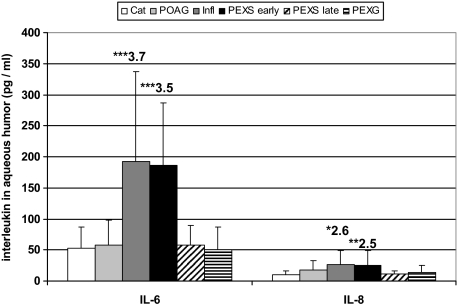
Pseudoexfoliation (PEX) syndrome, which is an age-related, generalized elastotic matrix process, currently represents the most common identifiable risk factor for open-angle glaucoma. Dysregulated expression of proinflammatory cytokines has been implicated in the initiation of various fibrotic disorders and in the pathophysiology of glaucoma. Here we investigated the presence, expression, regulation, and functional significance of proinflammatory cytokines in eyes with early and late stages of PEX syndrome/glaucoma in comparison with normal and glaucomatous control eyes using multiplex bead analysis, immunoassays, real-time PCR, Western blotting, immunohistochemistry, and cell culture models. Early stages of PEX syndrome were characterized by approximately threefold (P < 0.005) elevated interleukin (IL)-6 and IL-8 levels in the aqueous humor and a concomitant approximately twofold (P < 0.001) increase in mRNA expression levels in anterior segment tissues as compared with controls. In contrast, late stages of PEX syndrome/glaucoma did not differ significantly from controls. IL-6, IL-6 receptor, and phospho-signal transducer and activator of transcription 3 could be mainly localized to walls of iris vessels and to the nonpigmented epithelium of ciliary processes. IL-6 and IL-8 were significantly up-regulated by ciliary epithelial cells in response to hypoxia or oxidative stress in vitro, whereas IL-6, but not IL-8, induced the expression of transforming growth factor-beta1 and elastic fiber proteins. These findings support a role for a stress-induced, spatially, and temporally restricted subclinical inflammation in the onset of the fibrotic matrix process characteristic of PEX syndrome/glaucoma.


Figures

Figure 1
Figure 1
IL-6 and IL-8 concentrations in human aqueous humor as measured by multiplex bead immunoassay. Aqueous IL-6 and IL-8 levels are increased in early stages of PEX syndrome, whereas late stages and PEX glaucoma display no significant differences as compared with cataract or POAG. Results are expressed as mean ± SD together with the relative fold change as compared with cataract (Cat, cataract; POAG, primary open-angle glaucoma; Infl, resolved ocular inflammation; PEXS, PEX syndrome; PEXG, PEX glaucoma; n = 14 for each patient group; *P < 0.01, **P < 0.005, and ***P < 0.001).
Figure 2
Figure 2
IL-6 and total protein concentrations in human aqueous humor as determined by enzyme-linked immunosorbent assay and Bradford assay, respectively. A: Aqueous IL-6 levels are increased in early stages of PEX syndrome and in samples with previous ocular inflammation (n = 12 for each patient group). B: Total aqueous protein levels indicating function of the blood-aqueous barrier are increased in late stages of PEX without and with glaucoma and in eyes with previous inflammation. Results are expressed as mean ± SD together with the relative fold change as compared with cataract (Cat, cataract; POAG, primary open-angle glaucoma; Infl, resolved ocular inflammation; PEXS, PEX syndrome; PEXG, PEX glaucoma; n = 26 for each group; *P < 0.005 and **P < 0.001).
Figure 3
Figure 3
Quantitative determination of IL-6 and IL-8 mRNA expression levels in human ocular tissues using real-time PCR technology. A: IL-6 and IL-8 expression in various ocular tissues of normal human donor eyes. The expression levels were normalized against GAPDH, and the results are expressed as molecules IL per molecules GAPDH. The values represent mean values ± SD of four separate experiments. B and C: IL-6 and IL-8 mRNA expression in ciliary processes (B) and iris tissue (C) of PEX and control eyes; expression levels are increased in early stages of PEX syndrome. Data were normalized against GAPDH and are expressed as molecules interleukin per molecules GAPDH together with the relative fold change as compared with cataract. (PEXS, PEX syndrome; PEXG, PEX glaucoma; n = 6 for each patient group; *P < 0.001).
Figure 4
Figure 4
Western blot analysis of p-STAT3 and STAT3 in protein extracts from iris and ciliary processes of PEX and control eyes. A and B: Representative Western blot with 10 μg of iridal and ciliary total protein from two normal donors (control) and two patients with early and late stages of PEX syndrome (PEXS), respectively. Equal loading of samples was verified by immunodetection of β-actin. C: Intensities of specific immunoreactive bands were quantified by computerized densitometry, and signal intensity of control samples was set to 100%. Results are expressed as the ratio of p-STAT3 to STAT3, together with the relative fold change in early PEXS as compared with control (n = 5; *P < 0.005).
Figure 5
Figure 5
Immunofluorescence labeling of IL-6 in iris and ciliary process tissues of eyes with early and late stages of PEX syndrome (PEXS) and normal donor eyes (control). A, C, and E: Increased IL-6 immunopositivity (green fluorescence) is observed in blood vessel walls (arrows) and perivascular cells (arrowheads) of the iris stroma of an eye with early PEXS (age, 79 years) (C) as compared with late stage of PEXS without glaucoma (age, 81 years) (E) and normal control eye (age, 78 years) (A). B, D, and F: Increased IL-6 immunopositivity is observed in the nonpigmented epithelium (arrowheads) as well as in blood vessel walls (arrows) and stromal cells of ciliary processes of the same eyes with early PEXS (D) as compared with late stages of PEXS (F) and normal controls (B). (CE, ciliary epithelium; DM, dilator muscle; IPE, iris pigment epithelium; PEX, PEX material; ST, stroma; scale bar = 50 μm).
Figure 6
Figure 6
Immunofluorescence labeling of p-STAT3 in iris and ciliary process tissues of eyes with early and late stages of PEX syndrome (PEXS) and normal donor eyes (control). A, C, and E: Increased p-STAT3 immunopositivity (green fluorescence) is observed in stromal and perivascular cells (arrowheads) in the stroma and periphery of iris vessels (arrows) of an eye with early PEXS (age, 75 years) (C) as compared with late stage of PEXS without glaucoma (age, 78 years) (E) and normal control eye (age, 72 years) (A). B, D, and F: Increased p-STAT labeling is seen in stromal cells (arrowheads) of ciliary processes of the same eyes with early PEXS (D) as compared with late stages of PEXS (F) and normal controls (B). (AB, anterior iris border layer; CE, ciliary epithelium; PEX, PEX material; ST, stroma; scale bar = 50 μm).
Figure 7
Figure 7
Effect of hypoxia, oxidative stress, and TGF-β1 on the mRNA expression of IL-6 and IL-8 in cultured human NPE cells as determined by quantitative real-time PCR. The expression levels were normalized against GAPDH, and the results are expressed as molecules IL per molecules GAPDH together with the maximum fold change of induction in hypoxia- or H2O2-treated cells as compared with untreated control cells and the maximum fold change of suppression (−) by TGF-β1 in cells treated with H2O2 in the presence of TGF-β1 as compared with H2O2-treated cells. A: Expression levels of IL-6 and IL-8 are significantly induced by hypoxia (2% O2) as compared with untreated control cells. B: Expression levels of IL-6 and IL-8 are significantly up-regulated by oxidative stress (50 μmol/L H2O2) as compared with untreated control cells. TGF-β1 (5 ng/ml) significantly attenuated the H2O2-induced up-regulation of both IL-6 and IL-8 (ox. stress +TGF-β1) as compared with cells treated with H2O2 alone (ox. stress). The values represent mean values ± SD of three separate experiments; *P < 0.001 and **P < 0.0001.
Figure 8
Figure 8
Effect of IL-6 (10 ng/ml) on mRNA expression of ECM components and TGF-β isoforms in cultured human NPE cells as determined by quantitative real-time PCR. The expression levels were normalized against GAPDH, and the results are expressed as molecules per molecules GAPDH together with the maximum fold change in treated cells as compared with untreated control cells. A: IL-6 significantly induced the expression of fibrillin-1 and LTBP-1 but not fibulin-1 (×104). B: IL-6 significantly induced the expression of TGF-β1 (×103) and TGF-β2 (×104) but not TGF-β3 (×103). The values represent mean values ± SD of three separate experiments; *P < 0.001 and **P < 0.0001.

References

    1. Schlötzer-Schrehardt U, Naumann GOH. Ocular and systemic pseudoexfoliation syndrome. Am J Ophthalmol. 2006;141:921–937. - PubMed
    1. Ritch R, Schlötzer-Schrehardt U. Exfoliation syndrome. Surv Ophthalmol. 2001;45:265–315. - PubMed
    1. Ovodenko B, Rostagno A, Neubert TA, Shetty V, Thomas S, Yang A, Liebmann J, Ghiso J, Ritch R. Proteomic analysis of exfoliation deposits. Invest Ophthalmol Vis Sci. 2007;48:1447–1457. - PubMed
    1. Zenkel M, Kruse FE, Jünemann AG, Naumann GOH, Schlötzer-Schrehardt U. Deficiency of the extracellular chaperone clusterin in eyes with pseudoexfoliation syndrome may be implicated in the aggregation and deposition of pseudoexfoliative material. Invest Ophthalmol Vis Sci. 2006;47:1982–1990. - PubMed
    1. Schlötzer-Schrehardt U, Pasutto F, Sommer P, Hornstra I, Kruse FE, Naumann GO, Reis A, Zenkel M. Genotype-correlated expression of lysyl oxidase-like 1 in ocular tissues of patients with pseudoexfoliation syndrome/glaucoma and normal patients. Am J Pathol. 2008;173:1724–1735. - PMC - PubMed

Publication types

MeSH terms