. 2010 Jun;176(6):3085-97.
doi: 10.2353/ajpath.2010.090989. Epub 2010 Apr 15.

A rat model for choroidal neovascularization using subretinal lipid hydroperoxide injection

Affiliations

A rat model for choroidal neovascularization using subretinal lipid hydroperoxide injection

Takayuki Baba et al. Am J Pathol. 2010 Jun.

Abstract

The purpose of this study was to develop and characterize a rat model of choroidal neovascularization (CNV) as occurs in age-related macular degeneration. The lipid hydroperoxide 13(S)-hydroperoxy-9Z,11E-octadecadienoic acid (HpODE) is found in submacular Bruch's membrane in aged humans and has been reported to generate neovascularization in a rabbit model. Three weeks after a single subretinal injection of 30 microg of HpODE, eyes of Sprague-Dawley rats were harvested. Follow-up fluorescein angiography was done on other animals until 5 weeks postinjection. Histological studies, immunohistochemical staining, and flatmount choroids for CNV measurements were performed. In addition, we used murine neuronal, bovine endothelial, and human ARPE19 cells for testing the in vitro effects of HpODE. CNV developed in 85.7% of HpODE-injected eyes. The neovascular areas were significantly greater in HpODE-injected eyes compared with those in control eyes (P = 0.023). The CNV had maximum dye leakage at 3 weeks, which subsided by the 5th week. Histologically, CNV extended from the choriocapillaris into the subretinal space. ED1-positive macrophages were recruited to the site. In vitro assays demonstrated that only 30 ng/ml HpODE induced cell proliferation and migration of endothelial cells. HpODE-induced CNV was highly reproducible, and its natural course seems to be ideal for evaluating therapeutic modalities. Because HpODE has been isolated from aged humans, the HpODE-induced rat model seems to be a relevant experimental model for CNV in age-related macular degeneration.


Figures
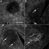
Figure 1
Figure 1
HpODE-induced CNV in flatmount choroids. Flatmount choroids at three weeks after injection were labeled with GSA-lectin to visualize the neovascularization. A: 1 μg of HpODE did not induce any demonstrable CNV formations. B and C: 5 μg (B) and 15 μg (C) of HpODE stimulated CNV development (arrows); however, the size of CNV formation was relatively small. D: 30 μg of HpODE stimulated well organized CNV formation of substantial size (arrow). Scale bars = 500 μm.
Figure 2
Figure 2
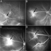
Fluorescein angiography of eye injected with 30 μg of HpODE. The time interval in each angiogram was almost the same, approximately 1.5 minutes after the dye injection. A: There was a small dye leakage at one week (arrow). The leakage area was greater at two weeks (B, arrow) and was greatest at three weeks (C, arrow). D: The leakage area decreased at five weeks (arrow).
Figure 3
Figure 3
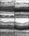
Histology after vehicle injection. There were no apparent changes in tissue morphology after vehicle injection at any time point. A: three days. B: one week. C: two weeks. D: three weeks. PAS and hematoxylin staining. Scale bars = 50 μm.
Figure 4
Figure 4
Sections through buffer (A–E) and HpODE injection sites at three days (F–J), one week (K–O), two weeks (P–T), and three weeks (U–Y) after injection. A: At three days after vehicle injection, there were no PAS-positive cells. B: Oil Red O staining is present only in photoreceptors. C: The retinal and choroidal vasculature is vWF-positive. There are only a few ED1-positive cells in inner retina (D) and ED2-positive cells in choroid (E). F: At three days after the HpODE injection, the sensory retina was still elevated, and there were many PAS-positive cells (paired arrows). G: Many subretinal cells and RPE cells contained lipids (paired arrows). Immunohistochemistry showed soluble vWF (H, asterisk) in the bleb and subretinal cells labeled with ED1 (I, arrow), whereas only cells in choroid were labeled with ED2 (J, arrowhead). One week after the HpODE injection, there were PAS-positive cells lined up along BrM (K, paired arrows), which contained lipids (L, paired arrows). Some of these cells were ED1-positive (N, paired arrows), and ED2 was present in cells of choroid (O, arrowhead). M: Blood vessels in inner retina, inner nuclear layer of retina, and choroid are labeled for vWf. At two weeks after the HpODE injection, the photoreceptors were degenerated (P, paired arrows). There were PAS-positive cells and material in degenerated retina and some lipids (Q, paired arrows). R: Blood vessels in retina and choroid are labled for vWf. Some cells in the retina were labeled with ED1 (S, paired arrows), and ED2-positive cells were restricted to choroid (T, arrowhead). By three weeks there was a robust subretinal CNV clearly labeled with vWF (W, open arrows). PAS-positive material extending horizontally in subretinal space (U, paired arrows), lipids (V, paired arrows), and some ED1-positive cells (X, paired arrows). Y: Only a fewED2-positive cells are present in choroid (arrowhead). Scale bars = 100 μm.
Figure 5
Figure 5
Histochemical staining after buffer (A–D) or HpODE (E–T) injection shown at high magnification. At three days after buffer injection, RPE cells were PAS-positive (A, paired arrows) but contained no lipids (B, paired arrows). There was no vWF (C, paired arrows) nor ED1-positive cells (D, paired arrows) in the subretinal space. E: At three days after the HpODE injection, there were many PAS-positive cells (paired arrows) in the subretinal space. F: Lipids were seen in these cells (paired arrows) and soluble vWF (G, paired arrows) was around the ED1-positive subretinal cells (H, paired arrows). One week after the HpODE injection there were PAS-positive cells lined up along BrM (I, paired arrows) with lipids (J, paired arrows). Some of these cells were ED1-positive (L, paired arrows). K: Some vWf+ structures, presumable deep retinal capillaries are present in the inner nuclear layer. M: At two weeks after the HpODE injection, the photoreceptors were degenerated. M and N: PAS-positive and lipid-laden cells were in the subretinal space (paired arrows). O: There is a vWf+ structure, presumable a deep retina capillary in what remains of the inner nuclear layer. P: Some subretinal cells labeled with ED1 (paired arrows). S: By three weeks there was a subretinal CNV formation clearly labeled with vWF (open arrows). PAS-positive material and cells are seen in subretinal space (Q, paired arrows), lipids (R, paired arrows), and some ED1-positive cells (T, paired arrows) above the CNV. Scale bars = 30 μm.
Figure 6
Figure 6
Extraction of lipids with chloroform-methanol. A: Oil Red O staining of lipids in RPE and monocytes (arrow, inset at higher magnification) at an injection site at seven days after injection of HpODE. B: A serial section to the one shown in A, which was extracted with chloroform/methanol and then stained with Oil Red O. The extraction process has removed all of the Oil Red O-stained lipids from the section in both RPE cells and monocytes (arrow, inset). C: PAS staining of a serial section. D: PAS staining of a serial section after chloroform/methanol extraction shows PAS-positive cells along BrM (arrow). No PAS-positive material is eliminated by the extraction process (arrow). Hematoxylin counterstaining in all. Scale bars = 20 μm.
Figure 7
Figure 7
CNV in JB4 sections. CNV formations (A, paired arrows) growing from choriocapillaris through BrM (B, arrowheads) into the subretinal space at three weeks after a 30-μg HpODE injection. C and D: A polymorphonuclear leukocyte is present in the CNV lumen (arrow). PAS and hematoxylin staining. Scale bar: 20 μm (A and C); 10 μm (B and D).
Figure 8
Figure 8
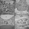
Ultrastructure after HpODE injection. A: At one week, the RPE cells contained many lipid droplets (arrowheads). The arrow indicates the large lipid droplet in the inset. The photoreceptors were degenerated at this site. B: At three weeks, there were fewer large lipid droplets in the RPE cell. C: Degeneration (arrowhead) and disorganization (bracket) of BrM were observed close to a lipid droplet (asterisk). D: The new vessels located just above BrM showed open lumens containing red blood cells (arrow). Scale bars: 2 μm (A); 4 μm (B); 500 nm (C); 4 μm (D).
Figure 9
Figure 9
HpODE dose-dependent cytotoxicity: photoreceptors (A), FBAECs (B), and ARPE19 cells (C). A–C: Although the lower concentrations of HpODE showed minimal cytotoxicity, 60 μg/ml HpODE was clearly cytotoxic. The photoreceptors, which started to die as early as four hours, were most vulnerable to high-dose HpODE (A). *P = 0.05.
Figure 10
Figure 10
HpODE effects on cell proliferation and migration. A: Proliferation assay, FBAECs. B: Proliferation assay, ARPE19 cells. C: Migration assay, FBAECs. D: Migration assay, ARPE19 cells. HpODE doses from 300 to 3 ng/ml produced significant cell proliferation of FBAECs and ARPE19 cells. A and B: 30 μg/ml HpODE showed a negative effect on cell proliferation because of its cytotoxicity. C and D: HpODE stimulated FBAEC migration at concentrations of 300 to 3 ng/ml (C) but did not stimulate a significant increase in ARPE19 cell migration (D). *P = 0.05.
Figure 11
Figure 11
Schematic diagram showing the proposed model of development of CNV induced by HpODE. HpODE injected subretinally (A) disperses and initiates the infiltration of macrophages and other inflammatory cells (B). Inflammatory cells and RPE phagocytose the HpODE (B). Progressive retinal degeneration begins in photoreceptor layer seven days after the HpODE injection (C). Proteolysis via the infiltrated cells degrades Bruch’s membrane (D). At seven to 14 days, the infiltrated and RPE cells stimulate migration and proliferation of choriocapillaris endothelial cells by secreting growth factors (E). The HpODE itself might stimulate endothelial cell proliferation and migration. Finally, CNV grows through Bruch’s membrane into the subretinal space (F).

References

    1. Ambati J, Ambati B, Yoo S, Ianchulev S, Adamis A. Age-related macular degeneration: etiology, pathogenesis, and therapeutic strategies. Surv Ophthalmol. 2003;48:257–293. - PubMed
    1. Green WR, Enger C. Age-related macular degeneration histopathologic studies. Ophthalmology. 1993;100:1519–1535. - PubMed
    1. Sarks SH. Aging and degeneration in the macular region: a clinico-pathological study. Br J Ophthal. 1976;60:324–341. - PMC - PubMed
    1. Ryan SJ. The development of an experimental model of subretinal neovascularization in disciform macular degeneration. Trans Am Ophthalmol Soc. 1979;77:707–745. - PMC - PubMed
    1. Goldberg MF. Editorial: Bruch’s membrane and vascular growth. Invest Ophthalmol. 1976;15:443–446. - PubMed

Publication types

MeSH terms

Substances