. 2010 Apr 16;328(5976):368-72.
doi: 10.1126/science.1182376.

Rapid diversification of cell signaling phenotypes by modular domain recombination

Affiliations

Rapid diversification of cell signaling phenotypes by modular domain recombination

Sergio G Peisajovich et al. Science. .

Abstract

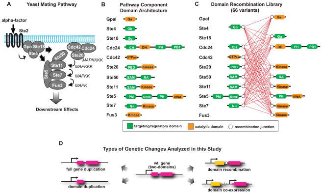
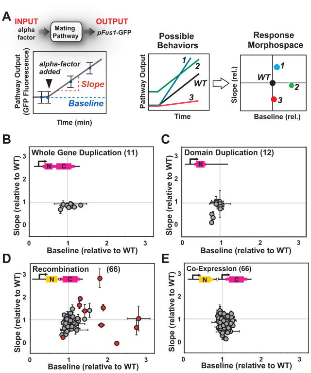
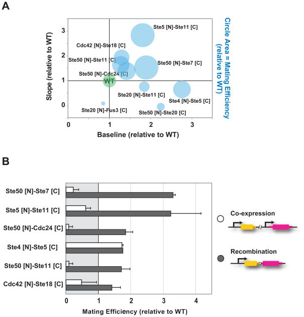
Cell signaling proteins are often modular, containing distinct catalytic and regulatory domains. Recombination of such biological modules has been proposed to be a major source of evolutionary innovation. We systematically analyzed the phenotypic diversity of a signaling response that results from domain recombination by using 11 proteins in the yeast mating pathway to construct a library of 66 chimeric domain recombinants. Domain recombination resulted in greater diversity in pathway response dynamics than did duplication of genes, of single domains, or of two unlinked domains. Domain recombination also led to changes in mating phenotype, including recombinants with increased mating efficiency over the wild type. Thus, novel linkages between preexisting domains may have a major role in the evolution of protein networks and novel phenotypic behaviors.


Conflict of interest statement

Competing Interests Statement

The authors declare no competing financial interests.

Figures

Fig. 1
Fig. 1. Design of the recombination library of protein domains belonging to the yeast mating pathway
A, the yeast mating pathway is activated by binding of the mating pheromone (α-factor) to the membrane receptor Ste2 (in “a” cells, or Ste3 in “α” cells), which causes the dissociation of the G protein alpha subunit (GpaI) from the G beta (Ste4) and gamma (Ste18) complex (20, 25). The scaffold protein Ste5 is then recruited to the membrane-localized Ste4, bringing along the MAPKKK Ste11, MAPKK Ste7 and MAPK Fus3. In addition, Ste11 interacts with the bridging protein Ste50, which by binding to the small GTPase Cdc42, positions Ste11 near its upstream activator, the PAK kinase Ste20 (26). Activated Ste11 phosphorylates Ste7, which in turn phosphorylates Fus3. The activated MAPK translocates to the nucleus where it phosphorylates a number of transcription factors, leading to changes in gene transcription, cell cycle progression and cell morphology and culminating in the fusion between “a” and “α” cells. B, domain architecture of the yeast mating signaling pathway components. Regulatory domains are shown in green, catalytic domains are shown in orange. Fully annotated domain maps are given in Fig. S8. C, domain recombination library: recombination junctions are depicted as white circles, all possible recombinations are shown as red connecting lines. D, possible evolutionary events analyzed in this work. Gene duplication, domain duplication, domain recombination and co-expression of two duplicated domains.
Fig. 2
Fig. 2. Recombination of protein domains results in diversification in signaling behaviors
A, mating pathway activation was measured by flow cytometry, using a GFP reporter under the control of a promoter (from the Fus1 gene) that responds to pathway activation, in an “a-type” ΔFar1 strain. Time course measurements of GFP fluorescence were done to calculate the baseline and slope of pathway activation, under conditions of linear pathway response. Baseline and slopes were then normalized relative to wild type values and the resulting values were plotted in pathway morphospace. B, gene duplications had minor effects on mating pathway response dynamics, with most values clustered around wild type. C, domain duplications had also minor effects on mating pathway response dynamics; duplication of Ste50 [N] (Ste50’s SAM domain), Ste5 [N] (which includes Ste5’s RING domain) and Ste11 [N] (Ste11’s SAM domain) are exceptions with low slopes and may act as dominant negative. D, domain recombination led to a diverse set of novel signaling behaviors. Recombination variants with dynamic behaviors most different from wild type and from the corresponding co-expressed N- and C-domain pair (Fig. S3) are shown in red. E, these behaviors could not be recapitulated by co-expression of the unlinked corresponding pairs of domains. Fluorescent values were measured in at least two independent experiments, each time in triplicates. Mean values +/− standard deviation (error bars) are depicted.
Fig. 3
Fig. 3. Domain recombination can lead to strains that mate more efficiently than wild type
A, mating efficiencies were measured for recombination variants with slope and baseline values that were substantially different from wild type (more than one standard deviation) and also different from the slope and baseline values of the corresponding co-expressed N- and C- pair (Fig. S3 and Fig. S4). Mating efficiencies of wild type and recombination variants are depicted as circles, with areas representing relative mating efficiencies. B, Comparison of domain recombination to co-expression of the corresponding domain pairs (wild type values are set to one). Ste50 SAM domain interacts with the Ste11 (MAPKKK) N-terminal SAM domain, facilitating the interaction of Ste11 with Ste20, its upstream activator. Thus, it is possible that, as an isolated domain, Ste50 [N] (as well as Ste11 [N]) act as dominant-negatives, competing for the interaction between the wild type proteins.
Fig. 4
Fig. 4. Mechanisms of recombination-derived changes in signaling behavior
A, activation of the mating pathway requires interactions between three multi-protein complexes: the membrane-bound G-protein complex, the membrane bound polarity complex and the MAPK complex. Seven novel connections between the three multiprotein complexes and three novel connections within an individual complex formed by the recombination variants analyzed. B, D and F, flow cytometry time course of pFus1-GFP for strains expressing Ste20 [N]-Ste11 [C], Ste5 [N]-Ste11 [C], and Ste50 [N]-Ste20 [C], respectively. C, E and G, fluorescence microscopy of strains expressing GFP-labeled Ste20 [N]-Ste11 [C], Ste5 [N]-Ste11 [C], and Ste50 [N]-Ste20 [C], respectively.

Comment in

  • Signalling: Making new connections.
    Wrighton KH. Wrighton KH. Nat Rev Genet. 2010 Jun;11(6):387. doi: 10.1038/nrg2801. Epub 2010 May 11. Nat Rev Genet. 2010. PMID: 20458345 No abstract available.
  • Cell signalling: Making new connections.
    Wrighton KH. Wrighton KH. Nat Rev Mol Cell Biol. 2010 Jun;11(6):386. doi: 10.1038/nrm2909. Epub 2010 May 12. Nat Rev Mol Cell Biol. 2010. PMID: 20461096 No abstract available.

Similar articles

Cited by

References

    1. Chothia C, Gough J, Vogel C, Teichmann SA. Science. 2003 Jun 13;300:1701. - PubMed
    1. Vogel C, Bashton M, Kerrison ND, Chothia C, Teichmann SA. Curr Opin Struct Biol. 2004 Apr;14:208. - PubMed
    1. Pawson T, Nash P. Science. 2003 Apr 18;300:445. - PubMed
    1. Voigt CA, Martinez C, Wang ZG, Mayo SL, Arnold FH. Nat Struct Biol. 2002 Jul;9:553. - PubMed
    1. Bashton M, Chothia C. Structure. 2007 Jan;15:85. - PubMed

Publication types

MeSH terms

Substances