. 2010 Jul;78(1):69-80.
doi: 10.1124/mol.110.063727. Epub 2010 Apr 15.

Identification of RanBP 9/10 as interacting partners for protein kinase C (PKC) gamma/delta and the D1 dopamine receptor: regulation of PKC-mediated receptor phosphorylation

Affiliations

Identification of RanBP 9/10 as interacting partners for protein kinase C (PKC) gamma/delta and the D1 dopamine receptor: regulation of PKC-mediated receptor phosphorylation

Elizabeth B Rex et al. Mol Pharmacol. 2010 Jul.

Abstract

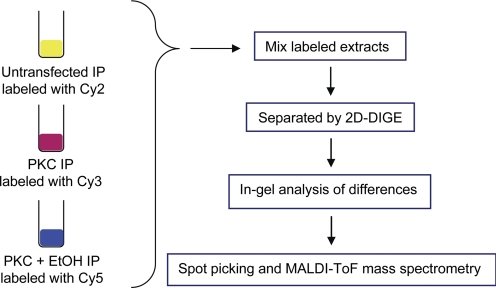
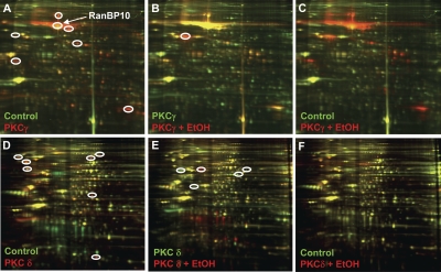
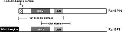
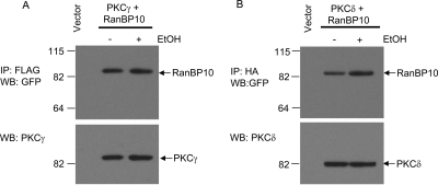
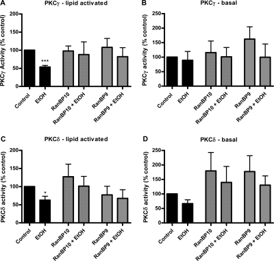
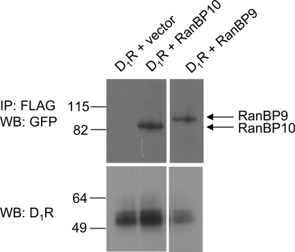
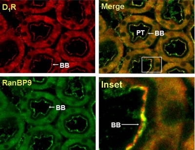
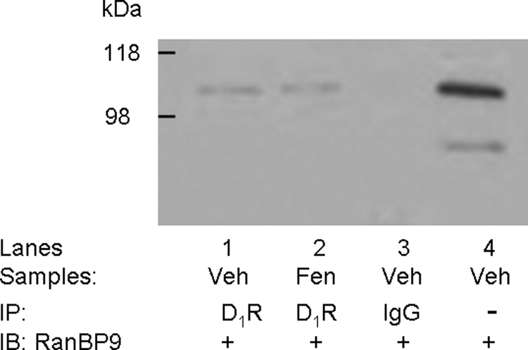
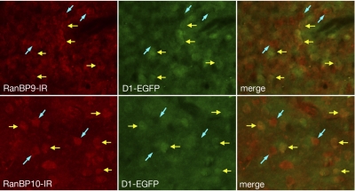
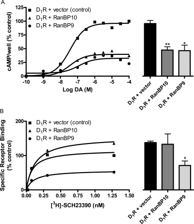
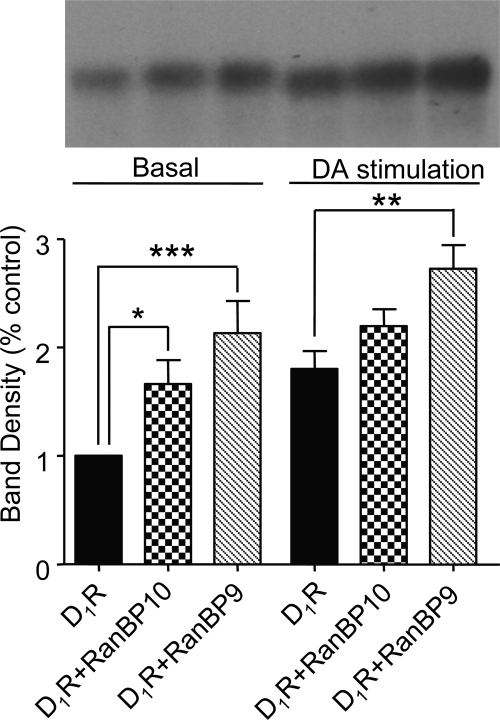
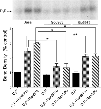
We reported previously that ethanol treatment regulates D(1) receptor phosphorylation and signaling in a protein kinase C (PKC) delta- and PKCgamma-dependent fashion by a mechanism that may involve PKC isozyme-specific interacting proteins. Using a PKC isozyme-specific coimmunoprecipitation approach coupled to mass spectrometry, we report the identification of RanBP9 and RanBP10 as novel interacting proteins for both PKCgamma and PKCdelta. Both RanBP9 and RanBP10 were found to specifically coimmunoprecipitate with both PKCgamma and PKCdelta; however, this association did not seem to mediate the ethanol regulation of the PKCs. It is noteworthy that the D(1) receptor was also found to specifically coimmunoprecipitate with RanBP9/10 from human embryonic kidney (HEK) 293T cells and with endogenous RanBP9 from rat kidney. RanBP9 and RanBP10 were also found to colocalize at the cellular level with the D(1) receptor in both kidney and brain tissue. Although overexpression of RanBP9 or RanBP10 in HEK293T cells did not seem to alter the kinase activities of either PKCdelta or PKCgamma, both RanBP proteins regulated D(1) receptor phosphorylation, signaling, and, in the case of RanBP9, expression. Specifically, overexpression of either RanBP9 or RanBP10 enhanced basal D(1) receptor phosphorylation, which was associated with attenuation of D(1) receptor-stimulated cAMP accumulation. Moreover, treatment of cells with select PKC inhibitors blocked the RanBP9/10-dependent increase in basal receptor phosphorylation, suggesting that phosphorylation of the receptor by PKC is regulated by RanBP9/10. These data support the idea that RanBP9 and RanBP10 may function as signaling integrators and dictate the efficient regulation of D(1) receptor signaling by PKCdelta and PKCgamma.


Figures

Fig. 1.
Fig. 1.
Experimental scheme for identifying candidate PKC-interacting proteins. FLAG-PKCγ or HA-PKCδ constructs were expressed in HEK293T cells, whereas untransfected cells served as controls. The cells were either untreated or pretreated with ethanol (100 mM/15 min), disrupted, and the PKC constructs were immunoprecipitated (IP) from the solubilized membrane fractions using antisera to either the FLAG or HA tags, as described under Materials and Methods. The immunoprecipitates were then labeled with CyDYE fluors as follows: untransfected cells, Cy2; PKC-transfected, Cy3; PKC-transfected plus ethanol treatment, Cy5. The labeled proteins were then resolved by 2D-gel electrophoresis and identified as described under Materials and Methods.
Fig. 2.
Fig. 2.
Identification of candidate PKC-interacting proteins. Each CyDYE-labeled PKC sample was simultaneously separated on a 2D-gel and scanned at specific wavelengths to reveal each of the CyDYE signals. Overlay of images reveal protein spots that are presumably specific (or nonspecific) to a given sample. Circled, protein spots selected for identification by mass spectrometry. A to C, PKCγ; D to F, PKCδ.
Fig. 3.
Fig. 3.
Structural organization of RanBP10 and RanBP9. The RanBP10 and RanBP9 proteins were aligned and compared. Conserved regions include a dual-specific kinase splA and ryanodine receptor domain, lissencephaly type-1-like homology domain, Ran binding protein domain, and guanine nucleotide exchange factor domain. RanBP10 contains a poly(proline/glutamine) tract at the amino terminus that is absent in RanBP9. [Adapted from Yudin D and Fainzilber M (2009) Ran on tracks–cytoplasmic roles for a nuclear regulator. J Cell Sci 122:587–593. Copyright © 2009 The Company of Biologists, Ltd. Used with permission.]
Fig. 4.
Fig. 4.
Association of RanBP10 with PKCγ and PKCδ. Coimmunoprecipitation and immunoblot analyses of RanBP10 with PKCγ (A) or PKCδ (B). HEK293T cells were transfected with empty vector, FLAG-PKCγ + RanBP10-GFP, or HA-PKCδ + RanBP10-GFP. PKCγ or PKCδ was immunoprecipitated from solubilized membrane fractions prepared from HEK293T cells that were either untreated or pretreated with ethanol (100 mM/15 min). As a control, the solubilized membrane fraction prepared from cells transfected with empty vector was incubated with either FLAG or HA beads. After SDS-PAGE, the gels were blotted with antisera directed to GFP (top) or to PKCγ or PKCδ. The experiment shown is representative of three independent experiments.
Fig. 5.
Fig. 5.
Effect of RanBP9 overexpression on ethanol-dependent attenuation of PKC activities. Epitope-tagged PKCs were immunoprecipitated from solubilized membrane fractions prepared from HEK293T cells coexpressing either PKCγ (A and B) or PKCδ (C and D) with vector, RanBP10, or RanBP9. The kinase activities of the isozyme-specific immunoprecipitates were directly assessed using an in vitro kinase assay as described under Materials and Methods. For each condition, the kinase activities were measured under basal or lipid-activated (plus phosphatidylserine and diacylglycerol) conditions in the presence or absence of 100 mM ethanol. Data are presented as the mean ± S.E.M. of at least three independent experiments. Results that are significantly different from the control groups are indicated as ∗, p < 0.05; ∗∗∗, p < 0.001, paired Student's t test.
Fig. 6.
Fig. 6.
RanBP10 and RanBP9 coimmunoprecipitate with the D1 receptor. The FLAG-D1 receptor was expressed in HEK293T cells either alone or with RanBP10-GFP or RanBP9-GFP. Membranes were prepared and solubilized followed by immunoprecipitation of the D1 receptor using anti-FLAG antisera as described under Materials and Methods. After resolution of the precipitates on SDS-PAGE, the gels were blotted with antisera directed to GFP (top) to detect RanB10 or RanBP9 or antisera directed to the D1 receptor (bottom). The experiment shown is representative of three independent experiments.
Fig. 7.
Fig. 7.
D1 receptor and RanBP9 colocalization in rat kidney. D1R (red) and RanBP9 (green) are expressed in the brush border (BB), apical membrane, and to a lesser extent observed in the cytoplasm of the proximal tubule (PT) (original magnification, 400×). The immunofluorescence images were acquired using Zeiss 510 confocal laser scanning microscope. Colocalization of the D1 receptor and RanBP9 is indicated by the yellow color in the merged image. The experiment shown is representative of three independent experiments.
Fig. 8.
Fig. 8.
Coimmunoprecipitation of the D1 receptor and RanBP9 endogenously expressed in kidney tissue. Immortalized renal proximal tubule cells (passage, ∼20, 80% confluent), made quiescent by incubating the cells for 2 h in medium without FBS, were treated with vehicle (Veh; sterile water, 10 min, lane 1) or a D1-like agonist, fenoldopam (Fen; 1 μM, 10 min, lane 2). Lysates of treated cells or IgG were immunoprecipitated (IP) and immunoblotted (IB) as shown. To determine the specificity of the bands, normal rabbit IgG was used for immunoprecipitation and served as the negative control (lane 3), and Veh-cell lysate (immunoblot, lane 4) was used as positive control. The studies were performed three times with similar results.
Fig. 9.
Fig. 9.
Cellular colocalization of the D1 receptor and RanBP9 in brain. Brain sections through the striatum from transgenic mice in which D1 receptors are marked with EGFP were processed for fluorescence immunohistochemical localization of RanBP9 (top) and RanBP10 (bottom). Immunoreactivity for both RanBP9 (RanBP-IR) and RanBP10 (RanBP-IR) colocalize with both D1-EGPF-positive striatal neurons (yellow arrows) and D1-EGFP-negative striatal neurons (blue arrows). Thus, both RanBP9 and RanBP10 seem to colocalize with striatal neurons expressing the D1 receptor.
Fig. 10.
Fig. 10.
Effect of RanBP10 and RanBP9 on D1 receptor expression and signaling. A, RanBP10 or RanBP9 was coexpressed with the D1 receptor in HEK293T cells followed by assessment of DA-stimulated cAMP accumulation. The curves on the left are representative experiments, whereas the histograms on the right represent averaged data from three experiments using 100 μM DA as the stimulus. Expression of either RanBP10 or RanBP9 decreased maximal DA-stimulated cAMP accumulation compared with control (D1 receptor + empty vector): Emax ± S.E.M. values for D1R + RanBP10 and D1R + RanBP9 were 47 ± 6 and 46 ± 10% of control, respectively. B, saturation radioligand binding experiments were performed on membranes prepared from control cells (D1 receptor + empty vector) or cells expressing the D1 receptor along with either RanBP10 or RanBP9. The curves on the left are representative experiments, whereas the histograms on the right represent averaged Bmax data from three experiments. RanBP9 coexpression reduced the D1 receptor Bmax value to 60 ± 14% of control. (∗, p < 0.05, ∗∗, p < 0.005, paired Student's t test).
Fig. 11.
Fig. 11.
RanBP10 and RanBP9 increase basal D1 receptor phosphorylation. In situ phosphorylation experiments were performed on HEK293T cells expressing the D1 receptor, D1 receptor + RanBP10, and D1 receptor + RanBP9 as described under Materials and Methods. Cells were incubated with media (control) or DA (10 μM) for 10 min. Top, autoradiogram of D1 receptor immunoprecipitates from a representative in situ phosphorylation assay. The lanes in the gel correspond to the bars at the bottom. Bottom, average values of band densities for each condition. The data are normalized as the percentage of control for each individual experiment. The histograms represent the mean ± S.E.M. from four independent experiments (∗, p < 0.05, ∗∗, p < 0.01, ∗∗∗, p < 0.001, analysis of variance followed by Bonferroni pair-wise comparisons).
Fig. 12.
Fig. 12.
The RanBP9 and RanBP10-induced increase of D1 receptor phosphorylation is blocked by PKC inhibitors. HEK293T cells expressing the D1 receptor, D1 receptor + RanBP10, and D1 receptor + RanBP9 were treated with the PKC inhibitors Gö6983 (10 μM) or Gö6976 (10 μM) for 60 min followed by the assessment of basal phosphorylation using an in situ phosphorylation assay as described in Fig. 11. Top, autoradiogram of D1 receptor immunoprecipitates from a representative in situ phosphorylation assay. The lanes in the gel correspond to the bars at the bottom. Bottom, average values of band densities for each condition. The data are normalized as the percentage of control for each individual experiment. The histograms represent the mean ± S.E.M. from three independent experiments (analysis of variance followed by Bonferroni pair-wise comparisons; ∗, p < 0.05, ∗∗, p = 0.056).

References

    1. Bermak JC, Li M, Bullock C, Zhou QY. (2001) Regulation of transport of the dopamine D1 receptor by a new membrane-associated ER protein. Nat Cell Biol 3:492–498 - PubMed
    1. Besson A, Wilson TL, Yong VW. (2002) The anchoring protein RACK1 links protein kinase Cepsilon to integrin beta chains. Requirements for adhesion and motility. J Biol Chem 277:22073–22084 - PubMed
    1. Csukai M, Chen CH, De Matteis MA, Mochly-Rosen D. (1997) The coatomer protein beta'-COP, a selective binding protein (RACK) for protein kinase Cepsilon. J Biol Chem 272:29200–29206 - PubMed
    1. Denti S, Sirri A, Cheli A, Rogge L, Innamorati G, Putignano S, Fabbri M, Pardi R, Bianchi E. (2004) RanBPM is a phosphoprotein that associates with the plasma membrane and interacts with the integrin LFA-1. J Biol Chem 279:13027–13034 - PubMed
    1. D'Souza MS, Ikegami A, Olsen CM, Duvauchelle CL. (2003) Chronic D1 agonist and ethanol coadministration facilitate ethanol-mediated behaviors. Pharmacol Biochem Behav 76:335–342 - PubMed

Publication types