. 2010 Jun 11;106(11):1722-30.
doi: 10.1161/CIRCRESAHA.109.214353. Epub 2010 Apr 15.

Erythropoietin activates mitochondrial biogenesis and couples red cell mass to mitochondrial mass in the heart

Affiliations

Erythropoietin activates mitochondrial biogenesis and couples red cell mass to mitochondrial mass in the heart

Martha S Carraway et al. Circ Res. .

Abstract

Rationale: Erythropoietin (EPO) is often administered to cardiac patients with anemia, particularly from chronic kidney disease, and stimulation of erythropoiesis may stabilize left ventricular and renal function by recruiting protective effects beyond the correction of anemia.

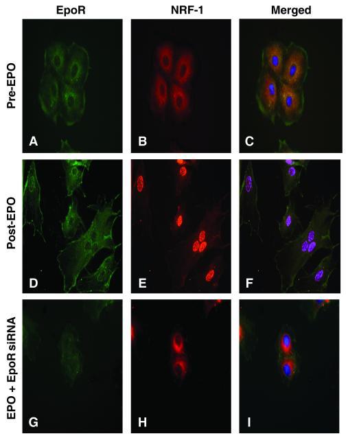
Objective: We examined the hypothesis that EPO receptor (EpoR) ligand-binding, which activates endothelial NO synthase (eNOS), regulates the prosurvival program of mitochondrial biogenesis in the heart.

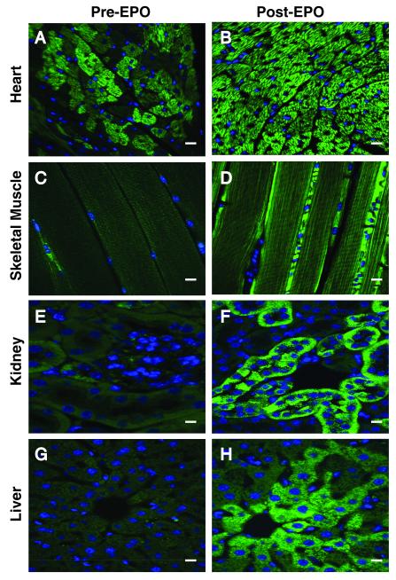
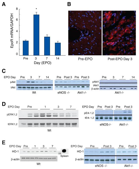
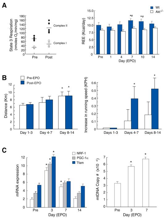
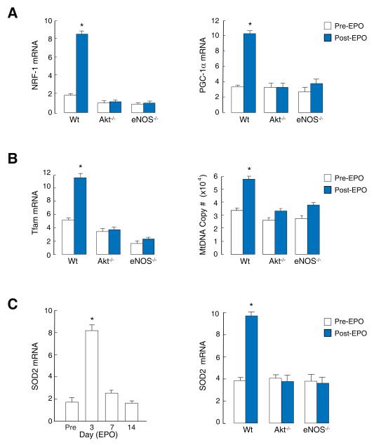
Methods and results: We investigated the effects of EPO on mitochondrial biogenesis over 14 days in healthy mice. Mice expressing a mitochondrial green fluorescent protein reporter construct demonstrated sharp increases in myocardial mitochondrial density after 3 days of EPO administration that peaked at 7 days and surpassed hepatic or renal effects and anteceded significant increases in blood hemoglobin content. Quantitatively, in wild-type mice, complex II activity, state 3 respiration, and mtDNA copy number increased significantly; also, resting energy expenditure and natural running speed improved, with no evidence of an increase in left ventricular mass index. Mechanistically, EPO activated cardiac mitochondrial biogenesis by enhancement of nuclear respiratory factor-1, PGC-1alpha (peroxisome proliferator-activated receptor gamma coactivator 1alpha), and mitochondrial transcription factor-A gene expression in wild-type but not in eNOS(-/-) or protein kinase B (Akt1)(-/-) mice. EpoR was required, because EpoR silencing in cardiomyocytes blocked EPO-mediated nuclear translocation of nuclear respiratory factor-1.

Conclusions: These findings support a new physiological and protective role for EPO, acting through its cell surface receptor and eNOS-Akt1 signal transduction, in matching cardiac mitochondrial mass to the convective O(2) transport capacity as erythrocyte mass expands.


Figures

Figure 1
Figure 1. Mitochondrial density and distribution in mitochondrial reporter mouse tissues
Reporter mice expressing a GPF-labeled mitochondrial localization sequence received EPO (4,000 U/kg/d) for three consecutive days and the sections were compared by fluorescence microscopy pre- and at day 3 post-EPO. Pre-EPO, green punctuate fluorescence, representing mitochondria, was present diffusely and staining was enhanced in sporadic clusters of cells. Post-EPO, the heart showed intense green fluorescence throughout the myocardium (A, B). More focal and less pronounced responses were found in skeletal muscle (C, D), kidney (E, F), and liver (G, H). Scale bars are 10 microns.
Figure 2
Figure 2. Cardiac EpoR expression and Akt1, Erk 1,2, and HO-1 activation by EPO
Cardiac EpoR transcript levels by real time RT-PCR at 3 days post-EPO administration to Wt mice. A. EpoR mRNA was detected pre-EPO and increased by post-EPO day 3 (*P < 0.05 vs. control; n=4 at each point). B. EpoR localization in mouse hearts pre- and post-EPO treatment (red fluorescence) with DAPI nuclear counterstain (blue). C. EPO activation of Akt1 in mouse heart. Phospho/total Akt was increased in Wt heart post-EPO day 3 and fell to control by day 14. Phospho/total Akt was unresponsive to EPO in eNOS -/- mice. Negative control are heart tissue from Akt1-/- mice probed with anti-pAkt and anti-Akt1 specific antibodies. D. Phospho/total Erk 1,2 activation in Wt, eNOS-/- and Akt1-/- hearts, pre- and post-EPO day 3. E. HO-1 protein expression in Wt, eNOS-/- and Akt1-/- mouse hearts.
Figure 3
Figure 3. REE, voluntary exercise, and mitochondrial biogenesis post-EPO administration in mice
A. Left panel: State 3 respiration in control and EPO-treated Wt mouse heart mitochondria with malate/glutamate (open circles) or succinate (closed circles) substrate (P < 0.05 pre vs. post EPO for State 3). Right panel: REE measured from oxygen uptake (VO2) and carbon dioxide production (VCO2) before and after EPO in Wt and Akt1-/- mice (*P < 0.05 vs. day 0, # P < 0.05 Wt vs. Akt1). REE failed to increase in Akt1-/- mice after EPO. B. Voluntary exercise on calibrated wheels. Data are averages for days 1-3, 4-7, and 8-14. Left panel: All mice were self-trained by increasing their running distance. Right panel: Running speed increases after EPO (* P< 0.05). C. Activation of cardiac mitochondrial biogenesis. NRF-1, PGC-1α and Tfam mRNA levels, and mtDNA copy number measured pre- and post-EPO in Wt control mouse hearts (*P<0.05 vs. control; n=4).
Figure 4
Figure 4. Activation of mitochondrial biogenesis by EPO is dependent on Akt and eNOS
A. Cardiac mRNA expression for NRF-1 and PGC-1α in Wt mice at day 3 post-EPO is absent in eNOS -/- and Akt1-/- mice. B. Increases in Tfam mRNA and mtDNA copy number in Wt mice at day 3 post-EPO is absent in eNOS -/- and Akt1-/- mice. C. SOD2 mRNA expression in Wt mouse heart pre- and post- EPO (left), and pre- and at post-EPO day 3 in Wt, eNOS -/- and Akt1-/- mice relative to GAPDH (right) (* P<0.05 vs. control; n=4).
Figure 5
Figure 5. EpoR requirement for EPO-mediated nuclear NRF-1 accumulation in cardiomyocytes
H9C2 rat cardiomyocytes were transfected with scrambled (Scr) or EpoR siRNA followed by EPO treatment (1000 U/ml). After 24h, fluorescence microscopy was performed using anti-NRF-1 (red) and EpoR (green). Panels A-C: Scr RNA Pre-EPO. Panels D-F: ScrRNA, Post-EPO; Panels G-I: Post-EPO + siRNA. Nuclear NRF-1 translocation by EPO requires EpoR expression.

Similar articles

Cited by

References

    1. Sawada K, Krantz SB, Kans JS, Dessyrpis EN, Sawyer S, Glick AD, Civin CL. Purification of human erythroid colony-forming units and demonstration of specific binding of erythropoietin. J Clin Invest. 1987;80:357–366. - PMC - PubMed
    1. Marrades RM, Roca J, Campistol JM, Diaz O, Barbera JA, Torregrosa JV, Masclans JR, Cobos A, Rodriguez-Roisin R, Wagner PD. Effects of erythropoietin on muscle O2 transport during exercise in patients with chronic renal failure. J Clin Invest. 1996;97:2092–2100. - PMC - PubMed
    1. Chong ZZ, Kang JQ, Maiese K. Erythropoietin is a novel vascular protectant through activation of Akt1 and mitochondrial modulation of cysteine proteases. Circulation. 2002;106:2973–2979. - PubMed
    1. Anagnostou A, Liu Z, Steiner M, Chin K, Lee ES, Kessimian N, Noguchi CT. Erythropoietin receptor mRNA expression in human endothelial cells. Proc Natl Acad Sci U S A. 1994;91:3974–3978. - PMC - PubMed
    1. Tsai PT, Ohab JJ, Kertesz N, Groszer M, Matter C, Gao J, Liu X, Wu H, Carmichael ST. A critical role of erythropoietin receptor in neurogenesis and post-stroke recovery. J Neurosci. 2006;26:1269–1274. - PMC - PubMed

Publication types

MeSH terms

Substances