Review
. 2010:2010:503906.
doi: 10.1155/2010/503906. Epub 2010 Apr 8.

There goes the neighborhood: pathological alterations in T-tubule morphology and consequences for cardiomyocyte Ca2+ handling

Affiliations
Review

There goes the neighborhood: pathological alterations in T-tubule morphology and consequences for cardiomyocyte Ca2+ handling

William E Louch et al. J Biomed Biotechnol. 2010.

Abstract

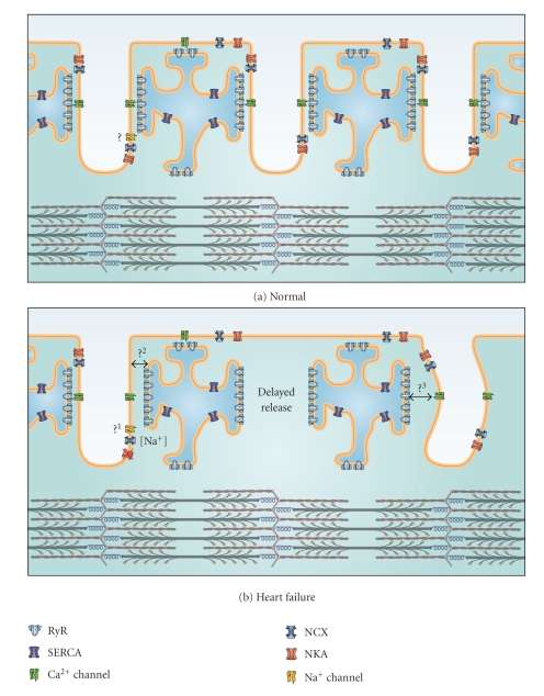
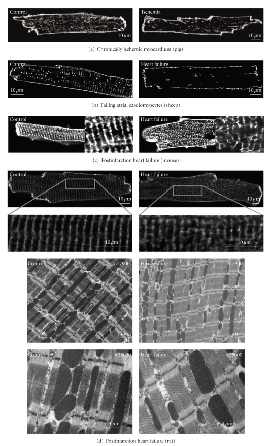
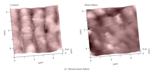
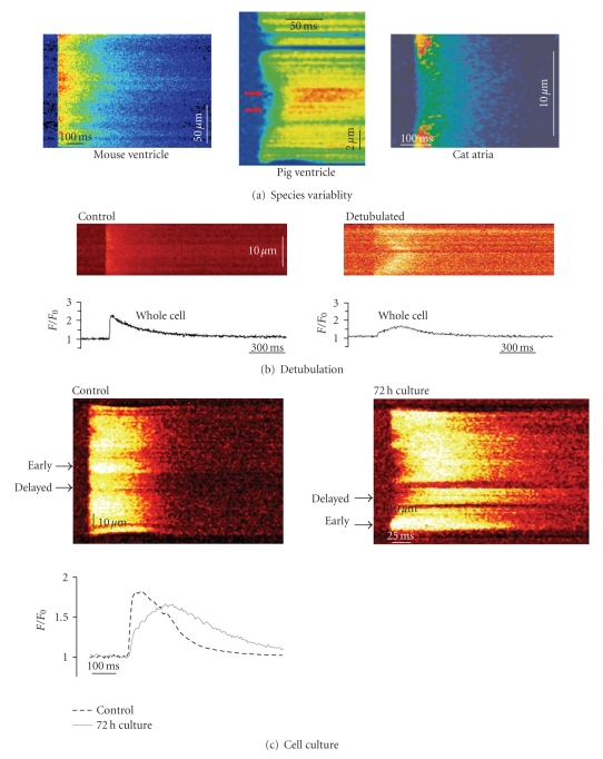
T-tubules are invaginations of the cardiomyocyte membrane into the cell interior which form a tortuous network. T-tubules provide proximity between the electrically excitable cell membrane and the sarcoplasmic reticulum, the main intracellular Ca2+ store. Tight coupling between the rapidly spreading action potential and Ca2+ release units in the SR membrane ensures synchronous Ca2+ release throughout the cardiomyocyte. This is a requirement for rapid and powerful contraction. In recent years, it has become clear that T-tubule structure and composition are altered in several pathological states which may importantly contribute to contractile defects in these conditions. In this review, we describe the "neighborhood" of proteins in the dyadic cleft which locally controls cardiomyocyte Ca2+ homeostasis and how alterations in T-tubule structure and composition may alter this neighborhood during heart failure, atrial fibrillation, and diabetic cardiomyopathy. Based on this evidence, we propose that T-tubules have the potential to serve as novel therapeutic targets.


Figures

Figure 1
Figure 1
Schematic representation of the dyadic neighborhood in normal and failing cells. (a) Excitation-contraction coupling occurs at functional junctions between Ca2+ channels in the T-tubules and ryanodine receptors in the SR. Depending on their localization, other proteins in the dyadic neighborhood such as SR Ca2+ ATPase (SERCA), NCX, NKA, and Na+ channels can also regulate Ca2+ homeostasis. Question mark: The positioning of the Na+ channel at the dyad is still controversial. (b) During heart failure, T-tubule loss and/or disorganization occurs leading to the formation of orphaned ryanodine receptors, which do not have apposing Ca2+ channels. Ca2+ release in these regions is delayed leading to slower and weaker contractions. Other putative alterations in the dyadic neighborhood are indicated by the question marks: (1) it is unclear whether the Na+ channel is present in the dyad of failing cardiomyocytes. Some experimental evidence suggests that the distance between the SR and T-tubule is increased in heart failure (2), while T-tubule disorganization may lead to dyadic clefts with variable width (3).
Figure 2
Figure 2
Examples of T-tubule alterations in various pathological states. Left panels show images from controls; right panels show images from (a) chronically ischemic pig myocardium (from [43]), (b) failing atrial ovine cardiomyocytes (from [40]), and post-infarction heart failure models in (c) mouse (from [44]) and (d) rat (from [45]). Panel (d) shows representative electron micrographs from control and failing rat hearts showing T-tubule disruption. (e) Scanning ion conductance microscope images from the surface of human nonfailing (control) and failing cardiomyocytes showing loss of T-tubular openings (from [46]). All figures are reproduced with permission.
Figure 2
Figure 2
Examples of T-tubule alterations in various pathological states. Left panels show images from controls; right panels show images from (a) chronically ischemic pig myocardium (from [43]), (b) failing atrial ovine cardiomyocytes (from [40]), and post-infarction heart failure models in (c) mouse (from [44]) and (d) rat (from [45]). Panel (d) shows representative electron micrographs from control and failing rat hearts showing T-tubule disruption. (e) Scanning ion conductance microscope images from the surface of human nonfailing (control) and failing cardiomyocytes showing loss of T-tubular openings (from [46]). All figures are reproduced with permission.
Figure 3
Figure 3
T-tubule density affects Ca2+ release synchrony. (a) Variable amount of T-tubules in different cell types affects the homogeneity of the Ca2+ transient, as illustrated in confocal line scans of myocytes from mouse ventricle ((a), unpublished data), pig ventricle ((b), from [43]), and cat atria ((c), from [49]). (b) Experimental loss of T-tubules using the detubulation technique causes delayed Ca2+ release in the centre of the cardiomyocyte (from [52]). (c) T-tubules are lost when cardiomyocytes are kept in culture resulting in dyssynchronous slowing of the Ca2+ transient (from [53]). All figures are reproduced with permission.
Figure 4
Figure 4
Dyssynchronous Ca2+ transients in failing cardiomyocytes. (a) Simultaneous imaging of T-tubules stained with di-8-ANEPPS and Ca2+ transients in fluo-4-AM loaded myocytes using confocal line scans. The position of the line scan is indicated as a vertical dotted line in T-tubule images, and T-tubules appear as horizontal lines in line-scan images. In myocytes from mice with congestive heart failure (CHF) following myocardial infarction, Ca2+ release was delayed in regions lacking T-tubules (right panel), but was synchronous in sham-operated controls (left panel), from [44]. (b) Regions of delayed Ca2+ release were also observed in cardiomyocytes from spontaneously hypertensive rats with heart failure (SHR/HF), but not in controls (Wistar-Kyoto (WKY) rats). From [47] Copyright (2006) National Academy of Sciences, USA. All figures are reproduced with permission.

References

    1. Brette F, Orchard C. Resurgence of cardiac T-tubule research. Physiology. 2007;22(3):167–173. - PubMed
    1. Forbes MS, Sperelakis N. The presence of transverse and axial tubules in the ventricular myocardium of embryonic and neonatal guinea pigs. Cell and Tissue Research. 1976;166(1):83–90. - PubMed
    1. Soeller C, Cannell MB. Examination of the transverse tubular system in living cardiac rat myocytes by 2-photon microscopy and digital image-processing techniques. Circulation Research. 1999;84(3):266–275. - PubMed
    1. Bers DM. Excitation-Contraction Coupling and Cardiac Contractile Force. Dordrecht, The Netherlands: Kluwer Academic Publishers; 2001.
    1. Hayashi T, Martone ME, Yu Z, et al. Three-dimensional electron microscopy reveals new details of membrane systems for Ca2+ signaling in the heart. Journal of Cell Science. 2009;122(7):1005–1013. - PMC - PubMed

Substances