

Healing of External Inflammatory Root Resorptions and Periapical Lesions without Surgical Treatment in an Operated Oblique Facial Cleft Case
- PMID: 20396455
- PMCID: PMC2853828
Healing of External Inflammatory Root Resorptions and Periapical Lesions without Surgical Treatment in an Operated Oblique Facial Cleft Case
Abstract
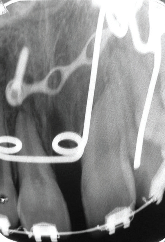
This paper describes an operated oblique facial cleft case with external inflammatory root resorption (EIRR) of the permanent maxillary left incisors and canine in a 12-year old patient. Due to the facial oblique cleft, the plastic surgery department operated on the patient five times and placed her on fixed orthodontic therapy. EIRR treatment of the maxillary left incisors and canine was performed using long-term calcium hydroxide therapy. The permanent root canal fillings of the lateral incisor and canine were performed using conventional gutta percha fillings. Because no sufficient apical barrier stop of the central incisor occurred, it was filled with Mineral Trioxide Aggregate (MTA); the canine crown fracture was restored using a carbon-covered fiberglass post and a light-cured composite resin. Examination after 42 months revealed good esthetics and no periapical pathology.
Keywords: External inflammatory root resorption; Oblique facial cleft; Tessier classification.
Figures


Similar articles
-
Mineral trioxide aggregate apical plug method for the treatment of nonvital immature permanent maxillary incisors: Three case reports.J Conserv Dent. 2012 Jan;15(1):73-6. doi: 10.4103/0972-0707.92611. J Conserv Dent. 2012. PMID: 22368340 Free PMC article.
-
Comparative evaluation of endodontic management of teeth with unformed apices with mineral trioxide aggregate and calcium hydroxide.J Dent Child (Chic). 2006 May-Aug;73(2):79-85. J Dent Child (Chic). 2006. PMID: 16948368
-
Single-visit endodontic treatment of mature teeth with chronic apical abscesses using mineral trioxide aggregate cement: a randomized clinical trial.BMC Oral Health. 2016 Aug 23;16(1):78. doi: 10.1186/s12903-016-0276-y. BMC Oral Health. 2016. PMID: 27553664 Free PMC article. Clinical Trial.
-
Root canal treatment of an immature dens invaginatus with apical periodontitis: a case report.J Dent Child (Chic). 2011 Jan-Apr;78(1):66-70. J Dent Child (Chic). 2011. PMID: 22041013
-
Conservative management of external root resorption after tooth reimplantation: a 3-year follow-up.Gen Dent. 2016 Jul-Aug;64(4):42-6. Gen Dent. 2016. PMID: 27367632
Cited by
-
The effect of retreatment procedure on the pH changes at the surface of root dentin using two different calcium hydroxide pastes.J Conserv Dent. 2012 Oct;15(4):346-50. doi: 10.4103/0972-0707.101899. J Conserv Dent. 2012. PMID: 23112482 Free PMC article.
References
-
- Kawamoto HJ. The kaleidoscopic world of rare craniofacial clefts:order out of chaos (Tessier classification) Clin Plast Surg. 1976;3:529–572. - PubMed
-
- Tessier P. Anatomical classification of facial, craniofacial and latero-facial clefts. J Maxillofac Surg. 1976;4:69–92. - PubMed
-
- Wenbin Z, Hanjiang W, Xiaoli C, Zhonglin L. Tessier 3 cleft with clinical anophthalmia: two case reports and a review of the literature. Cleft Palate Craniofac J. 2007;44:102–105. - PubMed
-
- George DI, Jr, Miller RL. Idiopathic resorption of teeth. A report of three cases. Am J Orthod. 1986;89:13–20. - PubMed
-
- Barnett F, Elfenbein L. Paget's disease of the mandible--a review and report of a case. Endod Dent Traumatol. 1985;1:39–42. - PubMed
LinkOut - more resources
Full Text Sources