. 2010 Apr 9;5(4):e10120.
doi: 10.1371/journal.pone.0010120.

CenH3/CID incorporation is not dependent on the chromatin assembly factor CHD1 in Drosophila

Affiliations

CenH3/CID incorporation is not dependent on the chromatin assembly factor CHD1 in Drosophila

Valerie Podhraski et al. PLoS One. .

Abstract

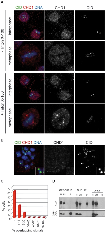
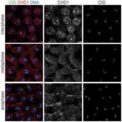
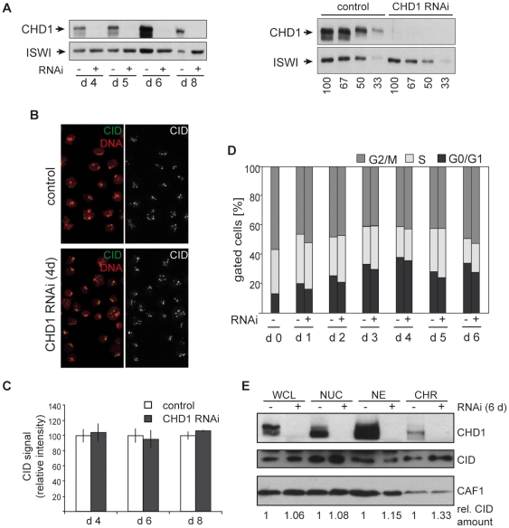
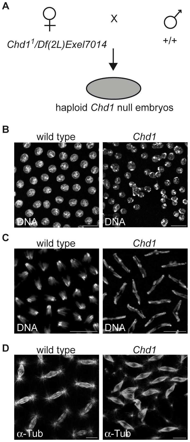
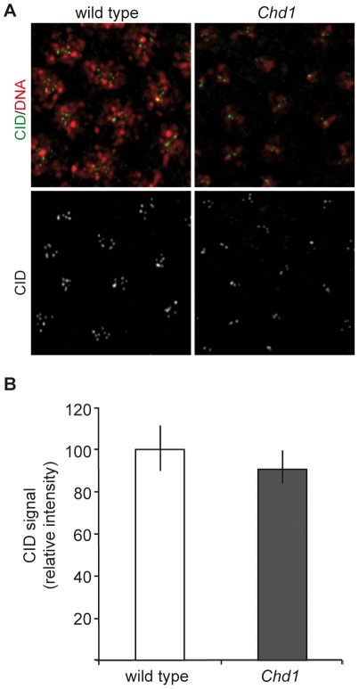
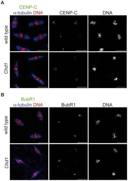
CHD1 is a SNF2-related ATPase that is required for the genome-wide incorporation of variant histone H3.3 in the paternal pronucleus as well as in transcriptionally active nuclei in Drosophila embryos. The S. pombe and vertebrate orthologs of CHD1 have been implicated in the assembly of the centromeric histone H3 variant CenH3(CENP-A), which occurs in a DNA replication-independent manner. Here, we examined whether CHD1 participates in the assembly of CenH3(CID) in Drosophila. In contrast to the findings in fission yeast and vertebrate cells, our evidence clearly argues against such a role for CHD1 in Drosophila. CHD1 does not localize to centromeres in either S2 cells or developing fly embryos. Down-regulation of CHD1 in S2 cells by RNAi reveals unchanged levels of CenH3(CID) at the centromeres. Most notably, ablation of functional CHD1 in Chd1 mutant fly embryos does not interfere with centromere and kinetochore assembly, as the levels and localization of CenH3(CID), CENP-C and BubR1 in the mutant embryos remain similar to those seen in wild-type embryos. These results indicate that Drosophila CHD1 has no direct function in the incorporation of the centromeric H3 variant CenH3(CID) into chromatin. Therefore, centromeric chromatin assembly may involve different mechanisms in different organisms.


Conflict of interest statement

Competing Interests: The authors have declared that no competing interests exist.

Figures

Figure 1
Figure 1. CHD1 is not present at centromeres in Drosophila S2 cells.
A) CHD1 displays nuclear staining during interphase and redistributes to the cytoplasm during mitosis. S2 cells stably expressing EGFP-tagged CenH3CID were treated (bottom) or not (top) with Triton X-100 before fixation to reduce the amount of soluble protein. Cells were stained with anti-CHD1 (red) and anti-GFP (green) antibodies. DNA is shown in blue. B) CHD1 is absent from chromosomes at metaphase. Spreads of metaphase chromosomes from EGFP-CenH3CID-expressing S2 cells were stained with antibodies against CHD1 (red) and GFP (green). C) Quantification of overlapping CHD1 and EGFP-CenH3CID signals. Percentages of overlapping signals per cell were calculated and plotted against the percentage of cells displaying similar ratios of overlap. D) CHD1 and EGFP-CenH3CID do not interact. Co-immunoprecipitations were performed on micrococcal nuclease treated S2 cell extracts with antibodies against GFP, CHD1 or protein A sepharose beads only. Aliquots of the input (IN) fraction, supernatant (SN) and eluted beads (B) were subjected to immunoblotting with anti-CID and anti-CHD1 antibodies.
Figure 2
Figure 2. CHD1 and CenH3CID do not colocalize in Drosophila embryos.
Embryos laid by transgenic female flies expressing EGFP-tagged CenH3CID were collected at 0–2 h after egg deposition and stained with antibodies against CHD1 (red) and GFP (green) to detect EGFP-CenH3CID. DNA was stained with DAPI (blue). Three syncytial embryos with interphase, metaphase and anaphase nuclei, respectively, are shown. CHD1 does not colocalize with centromeres at any cell cycle stage.
Figure 3
Figure 3. Cell cycle progression and CenH3CID loading are not affected by RNAi-mediated downregulation of CHD1.
A) Treatment of S2 cells with dsRNA targeting CHD1 for 4, 5, 6 and 8 days results in a decrease of CHD1 protein below detection limits. To control for equal loading western blots were incubated with an antibody against Drosophila ISWI (bottom). A serial dilution of control extracts was used to quantify CHD1 knockdown (right panel). B) CHD1 RNAi treated (4 days) and control cells were stained with anti-CenH3CID antibodies (green). DNA was counterstained with DAPI (red). C) Signal intensities of centromeric foci after staining with anti-CenH3CID antibodies were quantified as described in Materials and Methods. Error bars denote standard deviations of signals obtained from the following numbers of interphase nuclei: day 4 control and RNAi, n = 29; day 6 control, n = 168; day 6 RNAi, n = 148; day 8 control, n = 208; day 8 RNAi, n = 169. Signal intensities remained unchanged despite extensive CHD1 depletion. D) Cell cycle profiles of CHD1 RNAi treated (+) and untreated (−) control cells were determined by FACS analysis at different times (days 0–6) of dsRNA incubation. No significant changes in cell cycle progression patterns are detectable. E) Immunoblot analysis of cellular fractions of control and CHD1 RNAi treated cells with antibodies against CHD1 and CID. CAF-1 p55 was used as a loading control. WCL, whole cell lysate; NUC, nuclei; NE, high salt nuclear extract; CHR, chromatin core fraction (high salt insoluble fraction). The relative amounts of CID in the different fractions remain largely unaltered after CHD1-depletion.
Figure 4
Figure 4. A fraction of Drosophila Chd1 null embryos displays aberrant nuclear morphology and mitotic defects.
A) Schematic representation of the generation of Chd1 null embryos. B) The Chd1 mutation occasionally leads to the appearance of irregularly sized and arranged nuclei. C) Mitotic defects, such as lagging chromosomes and disorganized spindles (D), were observed in 15.6% of mutant embryos (n = 295) in contrast to only 1% (n = 209) of wild-type embryos. DNA was stained with DAPI (B, C), mitotic spindles were visualized by staining with an antibody against α-tubulin (D). Scale bars  = 10 µm.
Figure 5
Figure 5. CenH3CID loading is not compromised in Chd1 null embryos.
A) Embryos from wild-type and Chd1 null flies were stained with anti-CenH3CID antibodies (green). DNA was visualized with DAPI (red). B) Quantification of signal intensities of CenH3CID foci in wild-type and Chd1 null embryos (5 embryos each) as described in Material and Methods. CenH3CID signals have similar intensities indicating that CHD1 has no impact on CenH3CID loading.
Figure 6
Figure 6. Recruitment of kinetochore components is not disturbed in the absence of CHD1.
Wild-type and Chd1 null embryos were stained with antibodies against α-tubulin (blue) and either the inner kinetochore protein CENP-C (A) or the outer kinetochore protein BubR1 (B; both in green). DNA was counterstained with propidium iodide (red). Neither CENP-C nor BubR1 show apparent localization defects in the absence of CHD1. Scale bars  = 10 µm.

References

    1. Henikoff S, Dalal Y. Centromeric chromatin: what makes it unique? Curr Opin Genet Dev. 2005;15:177–184. - PubMed
    1. Haushalter KA, Kadonaga JT. Chromatin assembly by DNA-translocating motors. Nat Rev Mol Cell Biol. 2003;4:613–620. - PubMed
    1. Polo SE, Almouzni G. Chromatin assembly: a basic recipe with various flavours. Curr Opin Genet Dev. 2006;16:104–111. - PubMed
    1. Lusser A, Urwin DL, Kadonaga JT. Distinct activities of CHD1 and ACF in ATP-dependent chromatin assembly. Nat Struct Mol Biol. 2005;12:160–166. - PubMed
    1. Konev AY, Tribus M, Park SY, Podhraski V, Lim CY, et al. CHD1 motor protein is required for deposition of histone variant H3.3 into chromatin in vivo. Science. 2007;317:1087–1090. - PMC - PubMed

Publication types

LinkOut - more resources