Combined optical coherence tomography and intravascular ultrasound radio frequency data analysis for plaque characterization. Classification accuracy of human coronary plaques in vitro
- PMID: 20396951
- PMCID: PMC2991172
- DOI: 10.1007/s10554-010-9631-2
Combined optical coherence tomography and intravascular ultrasound radio frequency data analysis for plaque characterization. Classification accuracy of human coronary plaques in vitro
Abstract
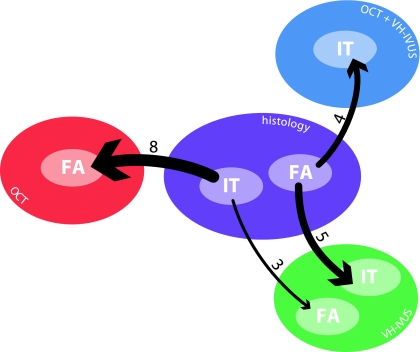
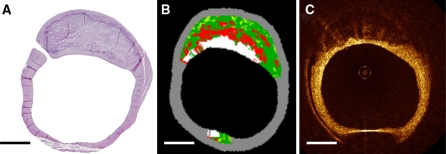
This study was performed to characterize coronary plaque types by optical coherence tomography (OCT) and intravascular ultrasound (IVUS) radiofrequency (RF) data analysis, and to investigate the possibility of error reduction by combining these techniques. Intracoronary imaging methods have greatly enhanced the diagnostic capabilities for the detection of high-risk atherosclerotic plaques. IVUS RF data analysis and OCT are two techniques focusing on plaque morphology and composition. Regions of interest were selected and imaged with OCT and IVUS in 50 sections, from 14 human coronary arteries, sectioned post-mortem from 14 hearts of patients dying of non-cardiovascular causes. Plaques were classified based on IVUS RF data analysis (VH-IVUS(TM)), OCT and the combination of those. Histology was the benchmark. Imaging with both modalities and coregistered histology was successful in 36 sections. OCT correctly classified 24; VH-IVUS 25, and VH-IVUS/OCT combined, 27 out of 36 cross-sections. Systematic misclassifications in OCT were intimal thickening classified as fibroatheroma in 8 cross-sections. Misclassifications in VH-IVUS were mainly fibroatheroma as intimal thickening in 5 cross-sections. Typical image artifacts were found to affect the interpretation of OCT data, misclassifying intimal thickening as fibroatheroma or thin-cap fibroatheroma. Adding VH-IVUS to OCT reduced the error rate in this study.
Figures


Similar articles
-
Direct Comparison of Virtual-Histology Intravascular Ultrasound and Optical Coherence Tomography Imaging for Identification of Thin-Cap Fibroatheroma.Circ Cardiovasc Imaging. 2015 Oct;8(10):e003487. doi: 10.1161/CIRCIMAGING.115.003487. Circ Cardiovasc Imaging. 2015. PMID: 26429760 Free PMC article.
-
Imaging assessment and accuracy in coronary artery autopsy: comparison of frequency-domain optical coherence tomography with intravascular ultrasound and histology.Int J Cardiovasc Imaging. 2019 Oct;35(10):1785-1790. doi: 10.1007/s10554-019-01639-0. Epub 2019 Jun 7. Int J Cardiovasc Imaging. 2019. PMID: 31175528
-
Additive Value of Integrated Backscatter IVUS for Detection of Vulnerable Plaque by Optical Frequency Domain Imaging: An Ex Vivo Autopsy Study of Human Coronary Arteries.JACC Cardiovasc Imaging. 2016 Feb;9(2):163-72. doi: 10.1016/j.jcmg.2015.07.011. Epub 2016 Jan 6. JACC Cardiovasc Imaging. 2016. PMID: 26777223
-
Intravascular ultrasound and optical coherence tomography imaging of coronary atherosclerosis.Int J Cardiovasc Imaging. 2016 Jan;32(1):189-200. doi: 10.1007/s10554-015-0701-3. Epub 2015 Jul 8. Int J Cardiovasc Imaging. 2016. PMID: 26153522 Review.
-
Clinical utility and significance of intravascular ultrasound and optical coherence tomography in guiding percutaneous coronary interventions.Circ J. 2015;79(1):24-33. doi: 10.1253/circj.CJ-14-1044. Epub 2014 Dec 19. Circ J. 2015. PMID: 25744628 Review.
Cited by
-
Complementary X-ray tomography techniques for histology-validated 3D imaging of soft and hard tissues using plaque-containing blood vessels as examples.Nat Protoc. 2014;9(6):1401-15. doi: 10.1038/nprot.2014.091. Epub 2014 May 22. Nat Protoc. 2014. PMID: 24853926
-
Plaque volume and plaque risk profile in diabetic vs. non-diabetic patients undergoing lipid-lowering therapy: a study based on 3D intravascular ultrasound and virtual histology.Cardiovasc Diabetol. 2017 Dec 7;16(1):156. doi: 10.1186/s12933-017-0637-0. Cardiovasc Diabetol. 2017. PMID: 29212544 Free PMC article. Clinical Trial.
-
In vivo and in situ cellular imaging full-field optical coherence tomography with a rigid endoscopic probe.Biomed Opt Express. 2011 Oct 1;2(10):2897-904. doi: 10.1364/BOE.2.002897. Epub 2011 Sep 29. Biomed Opt Express. 2011. PMID: 22025991 Free PMC article.
-
Direct Comparison of Virtual-Histology Intravascular Ultrasound and Optical Coherence Tomography Imaging for Identification of Thin-Cap Fibroatheroma.Circ Cardiovasc Imaging. 2015 Oct;8(10):e003487. doi: 10.1161/CIRCIMAGING.115.003487. Circ Cardiovasc Imaging. 2015. PMID: 26429760 Free PMC article.
-
Ultrasound and light: friend or foe? On the role of intravascular ultrasound in the era of optical coherence tomography.Int J Cardiovasc Imaging. 2011 Feb;27(2):209-14. doi: 10.1007/s10554-011-9797-2. Epub 2011 Feb 20. Int J Cardiovasc Imaging. 2011. PMID: 21337025 Free PMC article.
References
-
- Schaar JA, Muller JE, Falk E et al (2004) Terminology for high-risk and vulnerable coronary artery plaques. Report of a meeting on the vulnerable plaque, June 17 and 18, 2003, Santorini, Greece. Eur Heart J 25:1077–1082 - PubMed
Publication types
MeSH terms
LinkOut - more resources
Full Text Sources
Medical
Miscellaneous