. 2010:343:43-89.
doi: 10.1007/82_2010_37.

Picornaviruses

Affiliations

Picornaviruses

Tobias J Tuthill et al. Curr Top Microbiol Immunol. 2010.

Abstract

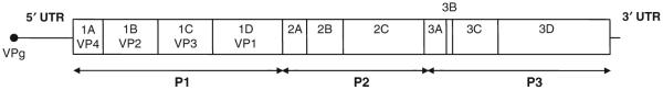
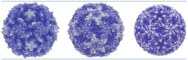
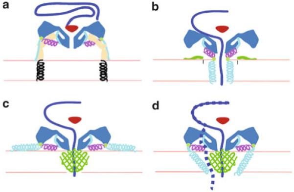
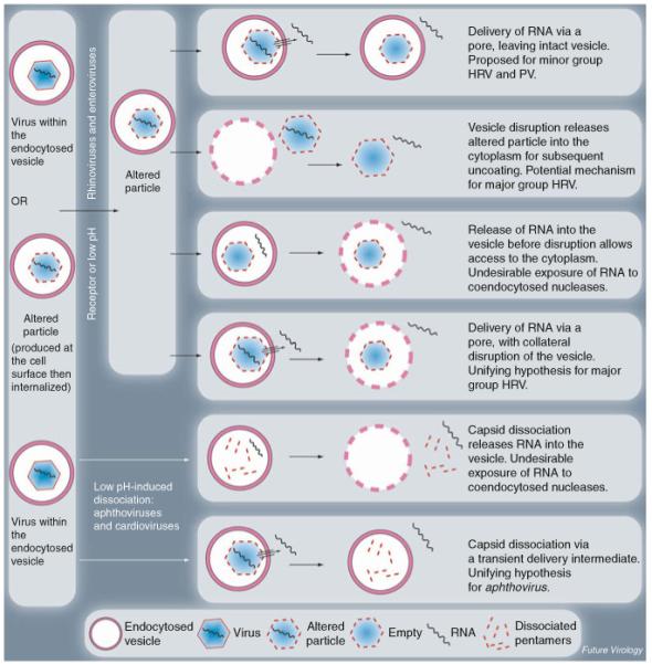
The picornavirus family consists of a large number of small RNA viruses, many of which are significant pathogens of humans and livestock. They are amongst the simplest of vertebrate viruses comprising a single stranded positive sense RNA genome within a T = 1 (quasi T = 3) icosahedral protein capsid of approximately 30 nm diameter. The structures of a number of picornaviruses have been determined at close to atomic resolution by X-ray crystallography. The structures of cell entry intermediate particles and complexes of virus particles with receptor molecules or antibodies have also been obtained by X-ray crystallography or at a lower resolution by cryo-electron microscopy. Many of the receptors used by different picornaviruses have been identified, and it is becoming increasingly apparent that many use co-receptors and alternative receptors to bind to and infect cells. However, the mechanisms by which these viruses release their genomes and transport them across a cellular membrane to gain access to the cytoplasm are still poorly understood. Indeed, detailed studies of cell entry mechanisms have been made only on a few members of the family, and it is yet to be established how broadly the results of these are applicable across the full spectrum of picornaviruses. Working models of the cell entry process are being developed for the best studied picornaviruses, the enteroviruses. These viruses maintain particle integrity throughout the infection process and function as genome delivery modules. However, there is currently no model to explain how viruses such as cardio- and aphthoviruses that appear to simply dissociate into subunits during uncoating deliver their genomes into the cytoplasm.


Figures

Fig. 1
Fig. 1
Picornavirus genome and polyprotein organization. The boxed area represents the single open reading frame (ORF) with untranslated regions (UTR) at each end. Translation and proteolytic processing produce primary products P1, P2, and P3. The P1 polyprotein is the capsid precursor and contains the structural proteins (VP1-4) found in the mature capsid. The P2 and P3 regions contain proteins involved in polyprotein processing, alteration of the host cell environment and replication of the viral RNA genome. The genome shown represents an enterovirus. Other genera contain subtle differences, for example, aphthoviruses contain an additional leader protein directly upstream of VP4, a small 2A protein and three copies of 3B which encode the genome-linked viral protein (VPg)
Fig. 2
Fig. 2
Picornavirus capsid structures. Radial depth cued images of picornavirus particles with a color gradient from innermost (dark blue) to outermost (white) surfaces. From left to right: poliovirus (enterovirus), 32 nm in diameter with five-pointed star shape at the fivefold axes, deep “canyon” surrounding the fivefold axes and three-bladed propeller at the threefold axes; Theiler’s murine encephalomyelitis virus (cardiovirus), 32 nm in diameter with extended star at the fivefold axes and surface depressions or “pits” spanning the twofold axes; foot-and-mouth disease virus (aphthovirus), 30 nm in diameter with relatively smooth surface. Images kindly produced by Hazel Levy
Fig. 3
Fig. 3
Working models for poliovirus entry. A cross section of a portion of the capsid is shown in dark blue, VP4 is green, and the N terminus of VP1 is cyan and magenta. (a) Native poliovirus binds its receptor, Pvr (ectodomains 1–3, tan; transmembrane domain, black helix), and at physiological temperature undergoes an irreversible change to the 135S particle. The path of egress of the N terminus of VP1 is shown. At this stage, the VP3 β tube (red) blocks an otherwise open channel along the fivefold axis. (b–d) Alternative models for the direct anchoring of the virus to the membrane via the N terminus of VP1 and formation of a transmembrane pore for RNA translocation. To accommodate the passage of RNA (purple), the VP3 β tube has shifted, and the channel has expanded, becoming continuous with a pore through the membrane. (b) Amphipathic helices at the N terminus of VP1 (cyan) may form a five-helix bundle close to the fivefold axis, which would require the magenta helix to dissociate from the body of the virus. Alternatively, VP4 may play a more central role in pore formation (c and d). In that case, VP1 may serve as a nonspecific membrane anchor (c) or participate directly in forming the pore (d). Recent studies have suggested an alternative path for release of the genome, from the base of the “canyon”, as indicated by the dashed line (d). Adapted from Bubeck et al. (2005a, b) with permission from the American Society for Microbiology
Fig. 4
Fig. 4
Possible scenarios for picornavirus structural changes and genome delivery to the cytosol during endocytosis. Adapted from Tuthill et al. (2007), with permission from Future Medicine

Similar articles

Cited by

References

    1. Acharya R, Fry E, Stuart D, Fox G, Rowlands D, Brown F. The three-dimensional structure of foot-and-mouth disease virus at 2.9 A resolution. Nature. 1989;337:709–716. - PubMed
    1. Akiyama SK. Integrins in cell adhesion and signaling. Hum Cell. 1996;9:181–186. - PubMed
    1. Andries K, Dewindt B, Snoeks J, Wouters L, Moereels H, Lewi PJ, Janssen PA. Two groups of rhinoviruses revealed by a panel of antiviral compounds present sequence divergence and differential pathogenicity. J Virol. 1990;64:1117–1123. - PMC - PubMed
    1. Aniento F, Emans N, Griffiths G, Gruenberg J. Cytoplasmic dynein-dependent vesicular transport from early to late endosomes. J Cell Biol. 1993;123:1373–1387. - PMC - PubMed
    1. Arnold E, Luo M, Vriend G, Rossmann MG, Palmenberg AC, Parks GD, Nicklin MJ, Wimmer E. Implications of the picornavirus capsid structure for polyprotein processing. Proc Natl Acad Sci USA. 1987;84:21–25. - PMC - PubMed

Publication types

LinkOut - more resources