Review
. 2010:342:189-209.
doi: 10.1007/82_2010_29.

Varicella-zoster virus T cell tropism and the pathogenesis of skin infection

Affiliations
Review

Varicella-zoster virus T cell tropism and the pathogenesis of skin infection

Ann M Arvin et al. Curr Top Microbiol Immunol. 2010.

Abstract

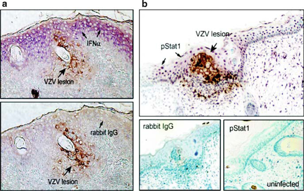
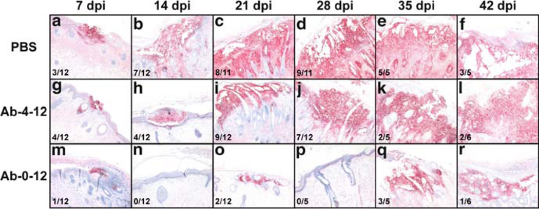
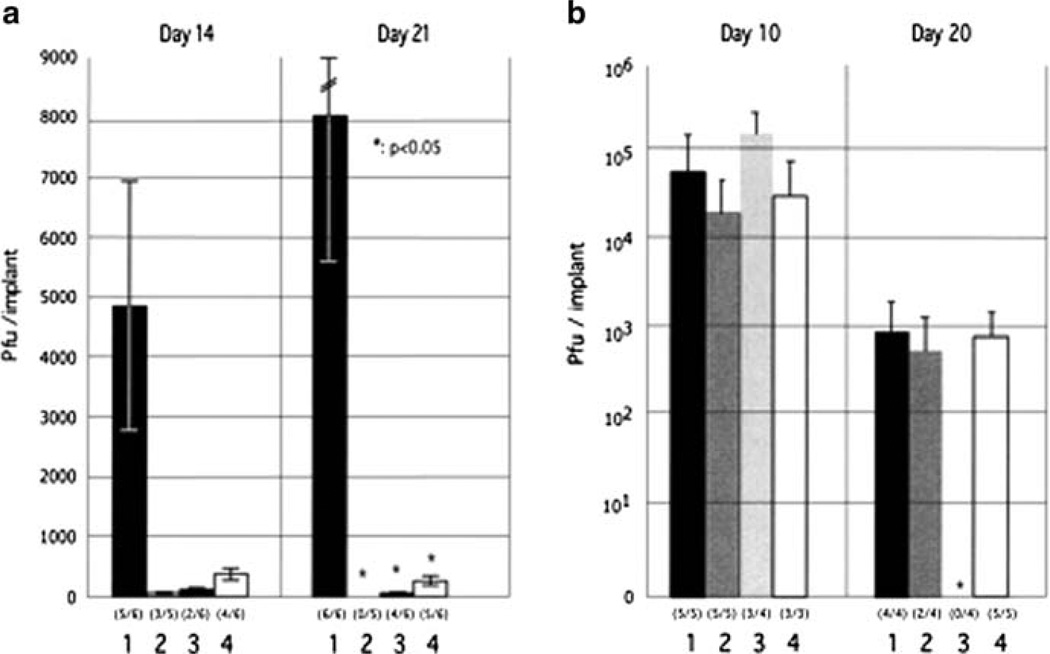
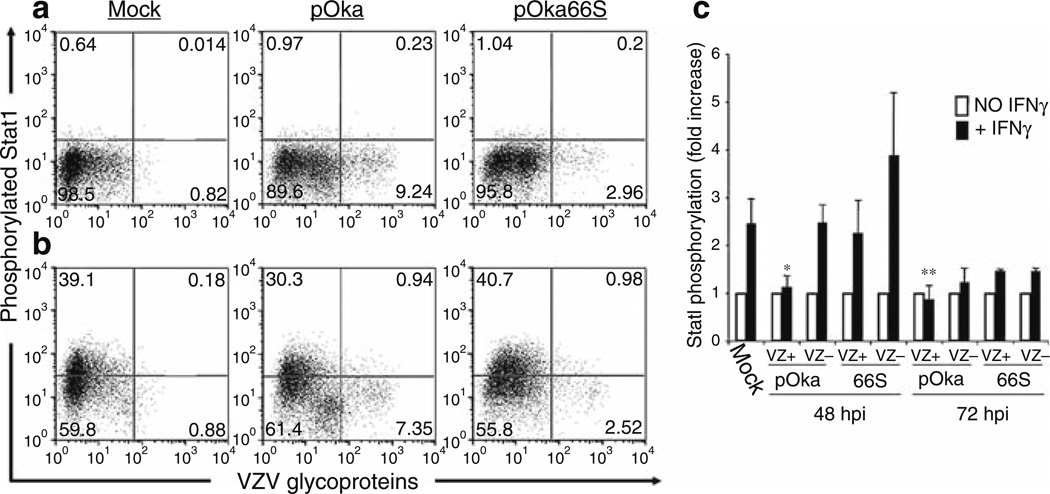
Varicella-zoster virus (VZV) is a medically important human alphaherpesvirus that causes varicella and zoster. VZV initiates primary infection by inoculation of the respiratory mucosa. In the course of primary infection, VZV establishes a life-long persistence in sensory ganglia; VZV reactivation from latency may result in zoster in healthy and immunocompromised patients. The VZV genome has at least 70 known or predicted open reading frames (ORFs), but understanding how these gene products function in virulence is difficult because VZV is a highly human-specific pathogen. We have addressed this obstacle by investigating VZV infection of human tissue xenografts in the severe combined immunodeficiency mouse model. In studies relevant to the pathogenesis of primary VZV infection, we have examined VZV infection of human T cell (thymus/liver) and skin xenografts. This work supports a new paradigm for VZV pathogenesis in which VZV T cell tropism provides a mechanism for delivering the virus to skin. We have also shown that VZV-infected T cells transfer VZV to neurons in sensory ganglia. The construction of infectious VZV recombinants that have deletions or targeted mutations of viral genes or their promoters and the evaluation of VZV mutants in T cell and skin xenografts has revealed determinants of VZV virulence that are important for T cell and skin tropism in vivo.


Figures

Fig. 1
Fig. 1
VZV infection of T cells in thymus/liver (T cell) xenografts in the SCID mouse model. On day 7 after infection, infected T cell xenografts were tested for VZV DNA by in situ hybridization; darkly stained cells indicate VZV DNA in T cells visualized at lower (left) and higher magnification (right; ×786). By electron microscopy on day 14 after infection, VZV nucleocapsids were present in the nuclei with most containing a dark VZV DNA core (large arrows); some empty capsids were also seen (small arrows). Virions were abundant in T cell nuclei, both individually and in clusters (arrowhead), also shown at higher magnification. Complete virions were found in the cytoplasm. Magnification and locations of cytoplasm (cyt) and nucleus (nuc) are as indicated. From Moffat et al. 1995 and Schaap et al. 2005; reproduced with permission
Fig. 2
Fig. 2
Role of cell fusion and polykaryocyte formation in VZV infection of skin xenografts. The upper panels show virion formation in skin xenografts infected with VZV (left panel) and kinase-defective ORF47 mutant (right panel) and examined at day 21 by staining with antibody to VZV gE and hematoxylin. Transmission microscopy demonstrated many intact viral particles in rOkainfected skin cells. Viral particles were incomplete and fewer in rOka47D-N infected skin cells. Arrows indicate polykaryocytes with dark staining of gE in plasma membranes of rOka-infected cells and persistent formation of polykaryocytes in skin infected with the ORF47 kinase mutant despite minimal gE localization to plasma membranes. As illustrated, virus-induced cell fusion, with the characteristic formation of polykaryocytes, supports VZV infection in skin but VZV infection does induce cell fusion. ORF47 kinase mutants do not infect T cells, suggesting that VZV T cell tropism depends upon efficient assembly and release of complete virions. (Adapted from Besser et al. 2004; figures reproduced with permission)
Fig. 3
Fig. 3
A model of the pathogenesis of primary and recurrent VZV infection. VZV infection is acquired by inoculation of mucosal epithelial cells via the respiratory route, transfer across epithelial layers allows infection of T cells in tonsils and other lymphoid tissue that comprise Waldeyer’s ring and allows transport to the skin via a T cell-associated viremia. Infection of skin produces the vesicular rash characteristic of varicella. VZV may reach sensory ganglia by T cell viremia or during skin infection, VZ virions may gain access to the sensory nerve cell body by retrograde axonal transport; lifelong latent infection is established in sensory ganglia in the course of primary VZV infection. Clinical reactivation of latent VZV results in herpes zoster, during which VZ particles gain access to skin via anterograde axonal transport. Adapted from Zerboni and Arvin 2008
Fig. 4
Fig. 4
Events in the pathogenesis of VZV skin infection. According to the model of primary VZV pathogenesis, T cells within the lymphoid tissues of Waldeyer’s ring become infected by VZV transfer following the initial inoculation of respiratory epithelial cells with the virus. Infected T cells enter the circulation and rapidly transport VZV to the skin, exiting from capillaries across endothelial cells by diapedesis, as occurs during the usual trafficking of T cells through tissues. VZV is then released by the infected T cells at skin sites of replication. The 10–21 day incubation period observed after exposure of naïve individuals to VZV is the interval required for VZV to overcome the innate IFN-α response mounted by epidermal cells and create a lesion that reaches the skin surface, IFN-α production by epidermal cells that surround those infected with VZV prevents a rapid, uncontrolled cell–cell spread that would otherwise incapacitate the host. Viremia may be amplified by trafficking of uninfected T cells through areas where skin lesions are forming. (Adapted from Ku et al. 2005; figure reproduced with permission)
Fig. 5
Fig. 5
IFN-α expression and Stat1 phosphorylation in VZV-infected and uninfected epidermal cells. Formalin-fixed, paraffin-embedded sections of VZV-infected skin xenografts were pretreated and double stained with anti-VZV IgG, detected with DAB (brown), and anti-IFNα or anti-pStat1 antibody detected with Vector VIP (purple). Sections are shown at magnification ×200. (a) Infected cells were identified by VZV protein (brown) expression. In double labeled skin sections, IFN-α (purple) expression was prominent in adjacent uninfected cells but not in VZV-infected cells (top) compared with sections stained with rabbit IgG as a control for IFN-α (bottom). (b) In double labeled sections, phosphorylated Stat1 (purple) was up-regulated in adjacent uninfected cells but absent in VZV-infected cells (top); pStat1 was not detected in uninfected skin (bottom right) compared with rabbit IgG control stain (bottom left). From Ku et al. 2004; reproduced with permission
Fig. 6
Fig. 6
The formation of lesions in VZV-infected human skin xenografts treated with the anti-gH mAb 206 for 0–12 days or 4–12 days post inoculation. Lesions in skin xenografts inoculated with pOka-infected HELF were identified by VZV gE expression, and were counterstained with hematoxylin. Representative lesions are shown at each timepoint: 7 dpi, A, G, M. 14 dpi, B, H, N. 21 dpi, C, I, O. 28 dpi, D, J, P. 35 dpi, E, K, Q. 42 dpi, F, L, R. 42 The total number of xenografts with lesions is shown in the lower 1 left of each panel. Lesions were identified in xenografts from the PBS (A–F) and Ab-4-12 (G–L) groups at all time points. Lesions were only observed in xenografts from the Ab-0-12 group (M–R) at 7, 21, 35 and 42 dpi. Representative uninfected xenografts are shown for the Ab-0-12 group at 14 and 28 dpi. Syncytia were seen in all lesions. Magnification: ×50. From Vleck et al. 2010, reproduced with permission
Fig. 7
Fig. 7
Replication of IE63 mutant viruses in skin and T cell xenografts in SCID-hu mice. Skin xenografts in SCID mice were injected with (left to right) rOKA, rOKA/ORF63rev[T171], rOKA/ ORF63rev[S181], or rOKA/ORF63rev[S185] having equivalent inoculum titers. Virus titers in skin xenografts were assessed after harvest at day 14 (A, left panel) and day 21 (A, right panel) after inoculation and were graphed as mean titers for xenografts that yielded infectious virus, with lines indicating standard errors. The number of xenografts from which infectious virus was recovered per number that were inoculated is given in parentheses below the horizontal axis. The P values were <0.05 when titers of rOka and each of the IE63 mutant viruses were compared at day 21. Replication of VZV recombinants in T cells was assessed at days 10 and 20 (B). Lines indicate the standard errors. From Baiker et al. 2004; reproduced with permission
Fig. 8
Fig. 8
Flow cytometric analysis of Stat1 phosphorylation in human tonsil T cells stimulated with IFN-γ. Column-purified human tonsil T cells were cocultured with VZV-infected HEL monolayers. After 48 h, cells were removed from the monolayer and either left unstimulated (a) or stimulated with recombinant human IFN-γfor 10 min at 37°C (b). Cells were fixed in paraformaldehyde, stained with antibodies and fluorescent conjugates to VZV proteins, permeabilized in methanol, and then stained with antibodies to phospho-Stat1 and CD3. FACS plots show antiphospho-Stat1 versus anti-VZV staining of uninfected (left plots), pOka-infected (middle plots), and pOka66S-infected (right plots) tonsil T cells (gated on CD3+ cells) without stimulation (a) or following stimulation with IFN-γ(b). (c) Data from T cells cultured for 48 or 72 h with infected HEL monolayers are shown as the average fold increase in phospho-Stat1 fluorescence intensity following IFN-γtreatment in VZV-infected and uninfected T cells for four independent experiments done after 48 h and two experiments combined for 72 h. One asterisk indicates a fold increase that is significantly different from that of uninfected cells from the same culture with P = 0.01, while two asterisks indicate a difference with P = 0.02. From Schaap et al. 2005; reproduced with permission
Fig. 9
Fig. 9
Lesion formation in skin xenografts infected with ORF10-to-ORF12 cluster gene mutants. Deparaffinized skin sections harvested at days 21 after infection were incubated with human polyclonal anti-VZV IgG and stained with biotinylated secondary antibody (magnification, ×10). Sections representative of skin xenografts infected with each virus are shown. Large lesions were observed for skin infected with POKA (a) and POKAΔ12 (f). Small lesions (arrows) restricted to the epidermal layer were observed for skin infected with POKAΔ11 (b), POKAΔ10/11 (c), and POKAΔ11/12 (d). POKAΔ10/11/12 (e) did not produce detectable skin lesions. The normal structures of the epidermis, the dermis, and the basement membrane that separates the epidermis from the underlying dermis are indicated in this section. From Che et al. 2008; reproduced with permission

Similar articles

Cited by

References

    1. Arvin AM. Varicella-zoster virus. In: Knipe DM, Howley P, editors. Fields’ virology. 4th edn. Philadelphia: Lippincott-Williams & Wilkins; 2001a. pp. 2731–2768.
    1. Arvin AM. Varicella vaccine: genesis, attenuation and efficacy. Virology. 2001b;284:153–158. - PubMed
    1. Arvin AM, Schaap AC, Ku C-C, Jones JO, Sommer M, Zerboni Z. Investigations of the molecular mechanisms of varicella-zoster virus pathogenesis. In: Sandri-Golden R, editor. The alphaherpesviruses. Horizon Press Inc: UK: 2006.
    1. Baiker A, Bagowski C, Ito H, Sommer M, Zerboni L, Fabell K, Hay J, Ruyechan W, Arvin AM. The immediate early 63 protein of varicella-zoster virus: analysis of functional domains required for replication in vitro and for T cell and skin tropism in the SCID hu model in vivo. J Virol. 2004;78:1181–1194. - PMC - PubMed
    1. Besser J, Sommer MH, Zerboni L, Bagowski C, Ito H, Moffat J, Ku C-C, Arvin AM. Differentiation of varicella-zoster virus ORF47 protein kinase and IE62 protein binding domains and their contributions to replication in human skin xenografts in the SCID-hu mouse. J Virol. 2003;77:5964–5974. - PMC - PubMed

Publication types