Spectroscopic and computational characterization of substrate-bound mouse cysteine dioxygenase: nature of the ferrous and ferric cysteine adducts and mechanistic implications
- PMID: 20397631
- PMCID: PMC2914100
- DOI: 10.1021/bi100189h
Spectroscopic and computational characterization of substrate-bound mouse cysteine dioxygenase: nature of the ferrous and ferric cysteine adducts and mechanistic implications
Abstract
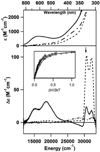
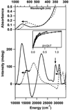
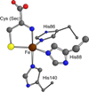
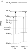
Cysteine dioxygenase (CDO) is a mononuclear non-heme Fe-dependent dioxygenase that catalyzes the initial step of oxidative cysteine catabolism. Its active site consists of an Fe(II) ion ligated by three histidine residues from the protein, an interesting variation on the more common 2-His-1-carboxylate motif found in many other non-heme Fe(II)-dependent enzymes. Multiple structural and kinetic studies of CDO have been carried out recently, resulting in a variety of proposed catalytic mechanisms; however, many open questions remain regarding the structure/function relationships of this vital enzyme. In this study, resting and substrate-bound forms of CDO in the Fe(II) and Fe(III) states, both of which are proposed to have important roles in this enzyme's catalytic mechanism, were characterized by utilizing various spectroscopic methods. The nature of the substrate/active site interactions was also explored using the cysteine analogue selenocysteine (Sec). Our electronic absorption, magnetic circular dichroism, and resonance Raman data exhibit features characteristic of direct S (or Se) ligation to both the high-spin Fe(II) and Fe(III) active site ions. The resulting Cys- (or Sec-) bound species were modeled and further characterized using density functional theory computations to generate experimentally validated geometric and electronic structure descriptions. Collectively, our results yield a more complete description of several catalytically relevant species and provide support for a reaction mechanism similar to that established for many structurally related 2-His-1-carboxylate Fe(II)-dependent dioxygenases.
Figures



Similar articles
-
Spectroscopic and Computational Investigation of the H155A Variant of Cysteine Dioxygenase: Geometric and Electronic Consequences of a Third-Sphere Amino Acid Substitution.Biochemistry. 2015 May 12;54(18):2874-84. doi: 10.1021/acs.biochem.5b00171. Epub 2015 May 1. Biochemistry. 2015. PMID: 25897562 Free PMC article.
-
Spectroscopic and computational characterization of the NO adduct of substrate-bound Fe(II) cysteine dioxygenase: insights into the mechanism of O2 activation.Biochemistry. 2013 Sep 3;52(35):6040-51. doi: 10.1021/bi400825c. Epub 2013 Aug 23. Biochemistry. 2013. PMID: 23906193 Free PMC article.
-
Spectroscopic and computational investigation of iron(III) cysteine dioxygenase: implications for the nature of the putative superoxo-Fe(III) intermediate.Biochemistry. 2014 Sep 16;53(36):5759-70. doi: 10.1021/bi500767x. Epub 2014 Aug 29. Biochemistry. 2014. PMID: 25093959 Free PMC article.
-
Cysteine dioxygenase: structure and mechanism.Chem Commun (Camb). 2007 Aug 28;(32):3338-49. doi: 10.1039/b702158e. Chem Commun (Camb). 2007. PMID: 18019494 Review.
-
Structure and function of atypically coordinated enzymatic mononuclear non-heme-Fe(II) centers.Coord Chem Rev. 2013 Jan 15;257(2):541-563. doi: 10.1016/j.ccr.2012.04.028. Coord Chem Rev. 2013. PMID: 24850951 Free PMC article. Review.
Cited by
-
Spectroscopic and Computational Comparisons of Thiolate-Ligated Ferric Nonheme Complexes to Cysteine Dioxygenase: Second-Sphere Effects on Substrate (Analogue) Positioning.Inorg Chem. 2019 Dec 16;58(24):16487-16499. doi: 10.1021/acs.inorgchem.9b02432. Epub 2019 Dec 2. Inorg Chem. 2019. PMID: 31789510 Free PMC article.
-
A Single DNA Point Mutation Leads to the Formation of a Cysteine-Tyrosine Crosslink in the Cysteine Dioxygenase from Bacillus subtilis.Biochemistry. 2023 Jun 20;62(12):1964-1975. doi: 10.1021/acs.biochem.3c00083. Epub 2023 Jun 7. Biochemistry. 2023. PMID: 37285547 Free PMC article.
-
Spectroscopic Investigation of Cysteamine Dioxygenase.Biochemistry. 2020 Jul 7;59(26):2450-2458. doi: 10.1021/acs.biochem.0c00267. Epub 2020 Jun 22. Biochemistry. 2020. PMID: 32510930 Free PMC article.
-
Spectroscopic analysis of the mammalian enzyme cysteine dioxygenase.Methods Enzymol. 2023;682:101-135. doi: 10.1016/bs.mie.2023.01.002. Epub 2023 Feb 15. Methods Enzymol. 2023. PMID: 36948699 Free PMC article.
-
Spectroscopic and Computational Investigation of the H155A Variant of Cysteine Dioxygenase: Geometric and Electronic Consequences of a Third-Sphere Amino Acid Substitution.Biochemistry. 2015 May 12;54(18):2874-84. doi: 10.1021/acs.biochem.5b00171. Epub 2015 May 1. Biochemistry. 2015. PMID: 25897562 Free PMC article.
References
-
- Yamaguchi K, Hosokawa Y. Cysteine dioxygenase. Methods Enzymol. 1987;143:395–403. - PubMed
-
- Lombardini JB, Singer TP, Boyer PD. Cysteine oxygenase. II. Studies on the mechanism of the reaction with 18oxygen. J. Biol. Chem. 1969;244:1172–1175. - PubMed
-
- Cooper AJL. Biochemistry of sulfur-containing amino acids. Annu. Rev. Biochem. 1983;52:187–222. - PubMed
-
- Stipanuk MH. Sulfur amino acid metabolism: pathways for production and removal of homocysteine and cysteine. Annu. Rev. Nutr. 2004;24:539–577. - PubMed
-
- Heafield MT, Fearn S, Steventon GB, Waring RH, Williams AC, Sturman SG. Plasma cysteine and sulphate levels in patients with motor neurone, Parkinson's and Alzheimer's disease. Neurosci. Lett. 1990;110:216–220. - PubMed
Publication types
MeSH terms
Substances
Grants and funding
LinkOut - more resources
Full Text Sources
Molecular Biology Databases

