Review
. 2010 Apr;8(4):545-56.
doi: 10.1586/erc.10.28.

Shear stress and plaque development

Affiliations
Review

Shear stress and plaque development

Saurabh S Dhawan et al. Expert Rev Cardiovasc Ther. 2010 Apr.

Abstract

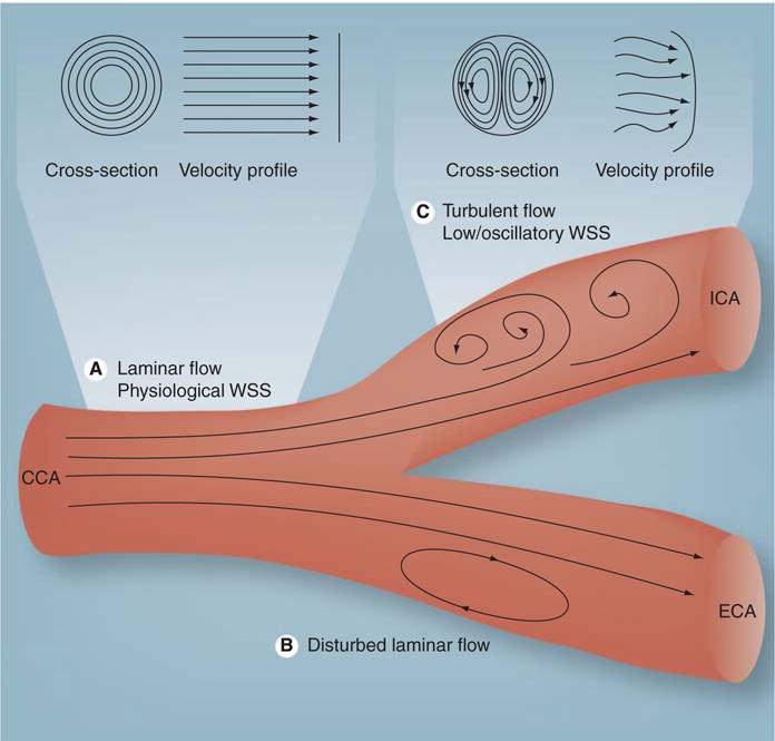
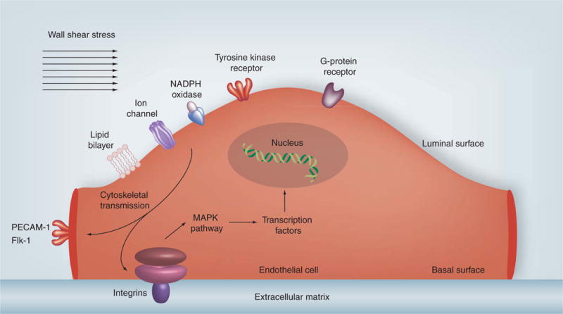
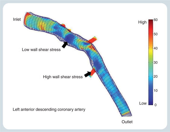
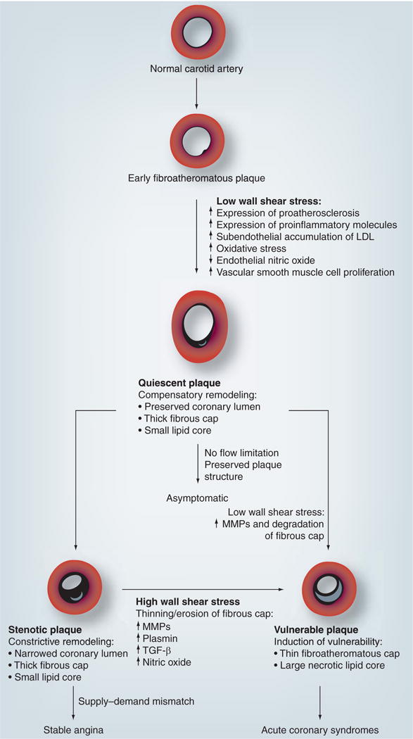
Although traditional cardiovascular risk factors 'prime the soil' for atherogenesis systemically, atherosclerosis primarily occurs in a site-specific manner with a predilection towards the inner wall of curvatures and outer wall of bifurcations with sparing of flow-dividers. Wall shear stress is a frictional force exerted parallel to the vessel wall that leads to alteration of the endothelial phenotype, endothelial cell signaling, gene and protein expression leading to a proinflammatory phenotype, reduced nitric oxide availability and disruption of the extracellular matrix, which in turn leads to plaque development. Clinical and experimental data are emerging that suggest the pathobiology associated with abnormal wall shear stress results in atherosclerotic plaque development and progression.


Conflict of interest statement

Financial & competing interests disclosure

The authors have no relevant affiliations or financial involvement with any organization or entity with a financial interest in or financial conflict with the subject matter or materials discussed in the manuscript. This includes employment, consultancies, honoraria, stock ownership or options, expert testimony, grants or patents received or pending, or royalties.

No writing assistance was utilized in the production of this manuscript.

Figures

Figure 1
Figure 1. Schematic diagram of the carotid bifurcation demonstrating different types of flow
(A) Laminar flow in which the layers of fluid move in a streamlined and concentric fashion to produce a conical velocity profile. (B) Disturbed laminar flow in which layers of fluid separate, flow in the reverse direction and then reattach to maintain forward flow. (C) Turbulent flow in which velocity of fluid varies continually, but the overall flow is in the forward direction albeit with a flatter velocity profile. CCA: Common carotid artery; ECA: External carotid artery; ICA: Internal carotid artery; WSS: Wall shear stress. Adapted from [100].
Figure 2
Figure 2. Effects of wall shear stress on inflammation and endothelial function
(A) Epifluorescence image of a whole mouse aortic mount showing the expression of ICAM-1 and VCAM-1 along the inner curvature of the arch and in the orifices of the arch vessels, areas that correspond to relatively lower values of mean wall shear stress (WSS). (B) Bar graphs depicting that partial ligation results in decreases in KLF-2 and eNOS, while increasing BMP4, ICAM-1 and VCAM-1 expression. (C) Impaired endothelium-dependent vasodilation as evident by lack of acetycholine-induced relaxation of arterial rings obtained from the left carotid artery of ApoE-knockout mice that underwent partial ligation and were fed a high-fat diet. (D) No impairment of endothelium-independent vasodilation as evident by complete relaxation induced by sodium nitroprusside. ACh: Acetylcholine; BMP: Bone morphogenic protein; eNOS: Endothelial nitric oxide synthase; KLF: Krüppel-like factor; LCA: Left carotid artery; PGF: Prostaglandin F; RCA: Right carotid artery; SNP: Sodium nitroprusside. Adapted from [37,46].
Figure 3
Figure 3. Schematic diagram broadly showing the regulation of transcription in the endothelial cells by wall shear stress
Wall shear stress is sensed on the luminal surface of endothelial cells by ionic channels, lipid layer, tyrosine kinase, G protein, NADPH oxidase and xanthine oxidase receptors that activate integrins on the basal surface of the endothelial cell or PECAM-1 and Flk-1 on the endothelial cell junctional surface via cytoskeletal transmission of shear forces. This initiates a downstream signaling cascade that activate MAPKs and other pathways to phosphorylate transcription factors, such as NF-κB and Krüppel-like factor 2, which regulate expression of atherogenic or atheroprotective genes. PECAM-1: Platelet endothelial cell adhesion molecule-1. Adapted from [47,98].
Figure 4
Figure 4
Wall shear stress profile of the left anterior descending coronary artery from a patient with nonobstructive coronary artery disease performed in our laboratory, outlining areas of low and high wall shear stress.
Figure 5
Figure 5. Development of atherosclerotic plaque and the role of wall shear stress in influencing plaque differentiation into quiescent, stenotic and vulnerable phenotypes
LDL: Low-density lipoprotein; MMP: Matrix metalloproteinase. Adapted from [47,98].

Similar articles

Cited by

References

    1. Ross R. Atherosclerosis – an inflammatory disease. N Engl J Med. 1999;340:11–26. - PubMed
    1. Van der Laan P, Reardon C, Getz G. Site specificity of atherosclerosis site-selective responses to atherosclerotic modulators. Arterioscler Thromb Vasc Biol. 2004:12–22. - PubMed
    1. Davies P. Flow-mediated endothelial mechanotransduction. Am Physiol Soc. 1995:51–60. - PMC - PubMed
    1. Nichols W, O’Rourke M. McDonald’s Blood Flow in Arteries: Theoretical, Experimental and Clinical Principles. 5th. Hodder Arnold Publications; London, UK: 2005.
    1. Slager CJ, Wentzel JJ, Gijsen FJH, et al. The role of shear stress in the generation of rupture-prone vulnerable plaques. Nat Clin Pract Cardiovasc Med. 2005;2:401–407. Excellent review of the pathobiology of plaque development and the generation of rupture-prone vulnerable plaques. - PubMed