. 2010 Jun;76(6):1461-82.
doi: 10.1111/j.1365-2958.2010.07151.x. Epub 2010 Apr 1.

The essential neutral sphingomyelinase is involved in the trafficking of the variant surface glycoprotein in the bloodstream form of Trypanosoma brucei

Affiliations
Free PMC article

The essential neutral sphingomyelinase is involved in the trafficking of the variant surface glycoprotein in the bloodstream form of Trypanosoma brucei

Simon A Young et al. Mol Microbiol. 2010 Jun.
Free PMC article

Abstract

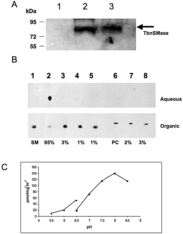
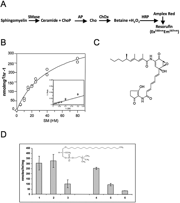
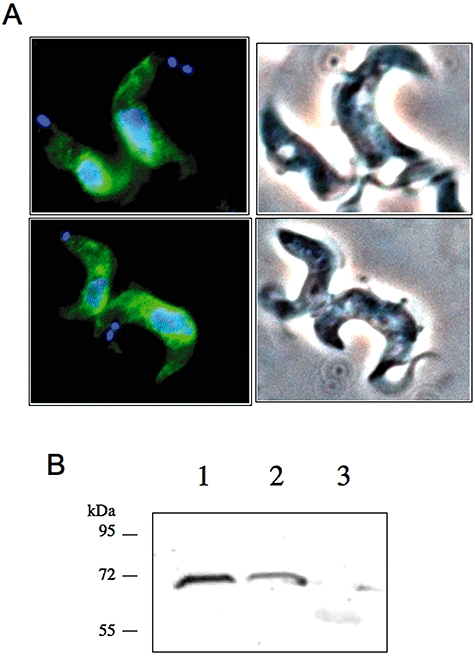
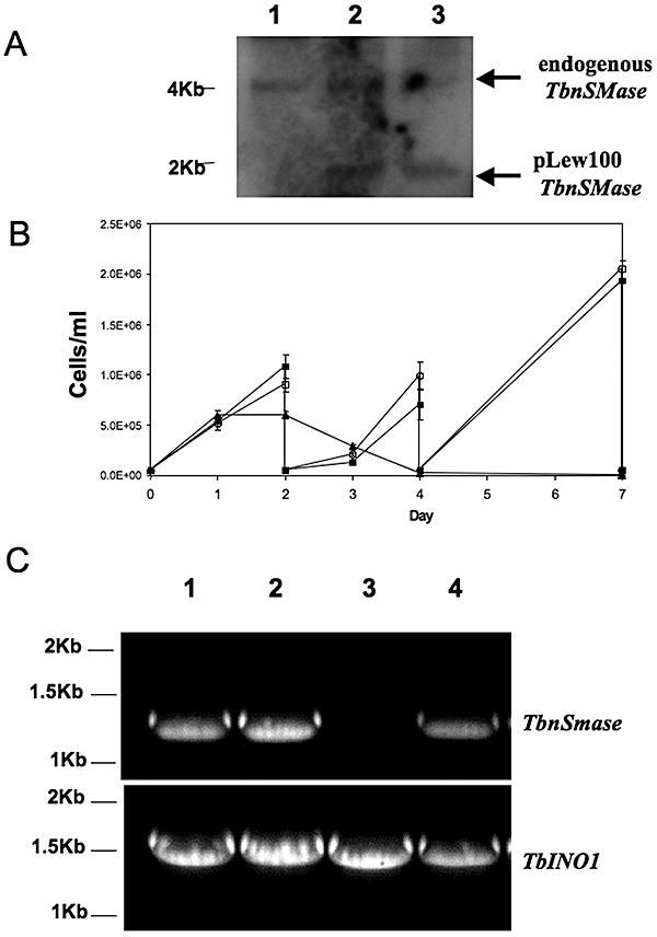
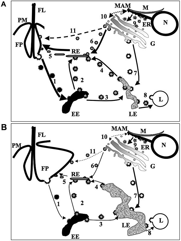
Sphingomyelin is the main sphingolipid in Trypanosoma brucei, the causative agent of African sleeping sickness. In vitro and in vivo characterization of the T. brucei neutral sphingomyelinase demonstrates that it is directly involved in sphingomyelin catabolism. Gene knockout studies in the bloodstream form of the parasite indicate that the neutral sphingomyelinase is essential for growth and survival, thus highlighting that the de novo biosynthesis of ceramide is unable to compensate for the loss of sphingomyelin catabolism. The phenotype of the conditional knockout has given new insights into the highly active endocytic and exocytic pathways in the bloodstream form of T. brucei. Hence, the formation of ceramide in the endoplasmic reticulum affects post-Golgi sorting and rate of deposition of newly synthesized GPI-anchored variant surface glycoprotein on the cell surface. This directly influences the corresponding rate of endocytosis, via the recycling endosomes, of pre-existing cell surface variant surface glycoprotein. The trypanosomes use this coupled endocytic and exocytic mechanism to maintain the cell density of its crucial variant surface glycoprotein protective coat. TbnSMase is therefore genetically validated as a drug target against African trypanosomes, and suggests that interfering with the endocytic transport of variant surface glycoprotein is a highly desirable strategy for drug development against African trypanosomasis.


Figures

Fig. 1
Fig. 1
Expression, purification and activity of recombinant TbnSMase. A. TbnSMase was cloned and expressed as an N-terminal GST fusion protein in C43 E. coli. Protein samples after cell disruption were separated on a 10% SDS-PAGE gel, transferred to membrane and detected with anti-GST antibody. Lane 1, soluble protein (supernatant from 100 000 g); lane 2, membrane fraction (pellet from 100 000 g); lane 3, insoluble fraction (pellet from 45 000 g). B. The substrate [3H]-sphingomyelin (SM) and the product [3H]-choline–phosphate of the TbnSMase assay were separated by phase partitioning (organic and aqueous respectively), analysed by HPTLC and detected by autoradiography as described in Experimental procedures. Lane 1, negative control, [3H]-SM with no membranes; lane 2, [3H]-SM with TbnSMase; lane 3, [3H]-SM with TbnSMase in presence of EDTA (5 mM); lane 4, negative control, [3H]-SM with non expressing TbnSMase membranes; lane 5, [3H]-SM with non-expressing TbnSMase membranes and EDTA (5 mM).; lane 6, [3H]-PC with no membranes; lane 7, [3H]-PC with TbnSMase; lane 8, [3H]-PC with TbnSMase in the presence of EDTA (5 mM). Percentages of substrate turnover are shown, as determined by densitometry (ImageJ software). C. TbnSMase activity was determined by end-point radioactive assay as a function of pH, using with MES buffer (filled triangles) or Bis-Tris Propane (filled squares) as described in Experimental procedures.
Fig. 2
Fig. 2
Activity of recombinant TbnSMase. A. Reaction catalysed by TbnSMase along with coupled the coupled Amplex red assay. AP, alkaline phosphatase; ChOx, choline oxidase; Cho-P, choline–phosphate; HRP, horseradish peroxidase. B. Determination of TbnSMase Michaelis-Menten constants for SM (inserts show Lineweaver–Burk plot). C. Structure of manumycin A, a commercially available nSMase inhibitor. D. Enzyme activity of TbnSMase in either washed bloodstream T. brucei membranes (lanes 1–3) or E. coli membranes expressing GST–TbnSMase (lanes 4–6), after pre-incubation with either nothing (lanes 1 and 4) or miltefosine (lanes 2 and 5) or edelfosine (lanes 3 and 6) in the presence of SM as substrate as described in Experimental procedures. Insert shows structure of edelfosine.
Fig. 3
Fig. 3
Localization of TbnSMase-HATi in bloodstream form T. brucei. A. Fixed TbnSMase-HATi cells were incubated with rat anti-HA antibody, and rabbit anti-rat FITC conjugated antibody and DNA stained with DAPI. B. Anti-HA Western analysis of fractions from differential centrifugation, 14 500 g pellet (1), 140 000 g pellet (2) and supernatant of 140 000 g pellet (3) of TbnSMase-HATi cells, prepared as described in the Experimental procedures.
Fig. 4
Fig. 4
TbnSMase is essential for the survival of bloodstream form Trypanosoma brucei in culture. A. Confirmation of genotype of T. brucei TbnSMase conditional double knockout cell line. Southern blot analysis of Pst1-digested genomic DNA (3 µg); the TbnSMase ORF probe shows allelic TbnSmase at 4 kb and the ectopic copy – TbnSMase-HATi at ∼2 kb; parental cells (lane 1); ΔnSMase:::PAC/TbnSMase-HATi (lane 2); ΔnSMase:::PACΔnSMase:::HYG/TbnSMase-HATi (lane 3). B. Growth curves of T. brucei parental cells (1 – filled squares) and TbnSmase conditional knockout cells grown in the presence (2 – empty circles) or absence (3 – filled triangles) of tetracycline. C. RT-PCR amplification of TbnSMase RNA transcripts from total RNA extracted from wild-type cells (lane 1) and TbnSMase conditional null mutants either grown in the presence (lane 2) or absence of tetracycline for 1 and 10 days (lanes 3 and 4 respectively). The upper panel shows RT-PCR products using primers specific for TbnSMase; the lower panel shows a loading control using (TbINO1) primers.
Fig. 5
Fig. 5
Mass spectrometric analyses of phospholipids. Choline-containing phospholipids extracted from the P100 fraction as described in Experimental procedures were analysed by ESI-MS/MS in positive ion mode using parent-ion scanning of the collision induced fragment for phosphorylcholine at 184 m/z in (A) parental cells. (B) TbnSMase cKO cells grown in the absence of tetracycline for 42 h. ESI-MS negative ion survey scan spectra of P100 lipid extracts from T. brucei bloodstream form cells. (C) Parental cells. (D) TbnSMase cKO cells grown in the absence of tetracycline for 42 h. Asterisks highlight ions, which are altered significantly in intensity, as described in the text
Fig. 6
Fig. 6
Morphological phenotype of the TbnSMase conditional knockout. A–D. Merged DAPI-fluorescence-DIC images of wild-type (A), and TbnSMase cKO cells grown in the absence of tetracycline for 42 h (B–D). E–J. Transmission electron micrographs taken of wild-type bloodstream T. brucei (E–G) and TbnSMase cKO cells grown in the absence of tetracycline for 42 h (H–J). Insert in (I) shows a close up of the plasma membrane, microtubles and the fussy VSG coat. FL, flagellum; FP, flagellar pocket; K, kinetoplast; M, mitochondria; G, glycosome; The asterisk in (J) indicates that enlarged concentric membrane structure may be part of the endosomal network. K–O. Wild-type (K) and TbnSMase cKO cells grown in the absence of tetracycline for 42 h (L–N) were incubated with FITC-BSA as described in Experimental procedures; images are merged FITC, DAPI-fluorescence and DIC. (O) Evaluation of the endocytic function of wild-type (1–3) and nSMase cKO (Tet-42h) (4–6) trypanosomes were assessed for the incidence of the FITC-BSA signal either solely in the endosomal region (1 and 4), or dual endosomal and flagellar pocket staining (2 and 5) or flagellar pocket staining only (3 and 6). P. FACS analysis of TbnSMase cKO cells grown in the absence of tetracycline for 0, 24 and 42 h. Cells were fixed, stained with propidium iodide and analysed by flow cytometry as described in Experimental procedures.
Fig. 8
Fig. 8
Phenotypic analysis of protein trafficking in the nSMase conditional knockout. A. Wild-type and TbnSMase cKO cells grown in the absence of tetracycline for 42 h were pulse labelled with [35S]-methionine for 1 h (lanes 1 and 3 respectively) and chased for further 2 h (lanes 2 and 4 respectively) prior to IP with anti-P67 as described in Experimental procedures. The resulting IP [35S]-proteins was separated on a 10% SDS-PAGE gel, prior to Coomassie blue staining (Fig. S6) and fluorography. The various maturation sizes of P67 are highlighted. B. Anti-trypanopain Western blot analysis of wild-type (lane 1) and TbnSMase cKO cells grown in the absence of tetracycline for 42 h (lane 2) shows incomplete endosomal processing. The maturation sizes of trypanopain are highlighted. C. Wild-type (lanes 1 and 3) and TbnSMase cKO cells grown in the absence of tetracycline for 42 h (lanes 2 and 4) were labelled with [35S]methionine for 1 h. Total [35S]-protein (lanes 1 and 2) or [35S]-sVSG (soluble VSG-obtained from the cell surface of the parasite) (lanes 3 and 4) prepared as described in Experimental procedures were separated on a 10% SDS-PAGE gel, and visualized either by Coomassie blue or fluorography. The black arrow (lane 1) refers to a protein that is no longer being synthesized in the TbnSMase cKO (-tet) cells (lane 2). D. Wild-type (lanes 1 and 2) and TbnSMase cKO cells grown in the absence of tetracycline for 42 h (lanes 3 and 4) were labelled with [3H]myristate for 1 h in the absence (lanes 1 and 4) or presence of cycloheximide (60 µg ml−1) (lanes 2 and 3) as described in Experimental procedures. Densitometry (ImageJ) of the [3H]myristate-mfVSG (membrane form VSG, processes an intact GPI-anchor) signals is normalized to 100% for the wild-type cells in the absence of cycloheximide. Lower panel shows the VSG portion of the gel stained with Coomassie blue as loading control.
Fig. 7
Fig. 7
Investigation of the endosomal network. Wild-type cells (A) and TbnSMase cKO cells grown in the absence of tetracycline for 42 h (B–D) probed with anti-trypanopain. Images are of TRITC, DAPI-fluorescence and DIC.
Fig. 9
Fig. 9
Schematic of the pathways involved in endocytosis and exocytosis in bloodstream form T. brucei: wild-type (A) and the nSMase cKO grown under permissive conditions (B). The difference in the width of the arrows in (A) and (B) depicts the relative changes in the flux for the same vesicular trafficking pathway in the wild-type cells compared with the nSMase cKO grown under permissive conditions. Dashed arrows indicate putative pathways. The enlarged flagellar pocket and late endosomes are representative of the ‘Big-Eye’ phenotype observed in the nSMase cKO grown under permissive conditions (B). Annotated cellular structures: EE, early endosomes; ER, endoplasmic reticulum; FP, flagellar pocket; G, Golgi; L, lysosome; LE, late endosomes; M, mitochondria; MAM, mitochondrion associated membrane; PM, plasma membrane; RE, recycling endosomes; FL, flagellum. The various types of vesicles are numbered as follows: 1. Endocytic clathrin-coated vesicles from flagellar pocket to early endosomes, carrying a mixture of endocytosed nutrients, including LDL particles and transferrin bound to GPI-anchored transferrin receptor, and recycling pre-existing cell surface GPI-anchored VSG, to be checked and undergoing myristate exchange. 2. Clathrin-coated vesicles from early endosomes to recycling endosomes carrying recycling pre-existing cell surface GPI-anchored VSG. 3. Clathrin-coated vesicles from early endosomes to late endosomes, carrying a mixture of endocytosed nutrients, including LDL particles and transferrin bound to GPI-anchored transferrin receptor. 4. Clathrin-coated vesicles from late endosomes to recycling endosomes, carrying the recycled GPI-anchored transferrin receptor, now free of transferrin, and newly synthesized membrane-bound protein destined for the flagellar pocket, not newly synthesized GPI-anchored proteins, i.e. VSG. 5. Non-clathrin-coated, sphingomylein-enriched recycling endosomes destined for the flagellar pocket, which contains recycling GPI-anchored transferrin receptor, recycling VSG and newly synthesized VSG. 6. Non-clathrin-coated, SM-enriched vesicles from the trans-Golgi cisterna to the recycling endosomes, containing newly synthesized VSG and possibly other GPI-anchored proteins, ultimately destined for the flagellar pocket and the cell surface. 7. Clathrin-coated vesicles from the Golgi to late endosomes, containing newly synthesized proteins which will be targeted to go either to the lysosome, i.e. p67 and trypanopain, or to go via the recycling endosomes to the flagellar pocket, i.e. cell surface proteins other than VSG. 8. Clathrin-coated vesicles from the late endosomes to the lysosome, containing a mixture of newly synthesized lysosomal proteins, i.e. p67 and trypanopain, and endocytosed nutrients, i.e. LDL particles and transferrin. 9. CopII vesicles from the ER to the Golgi, carrying all newly synthesized proteins destined for the endocytic pathway. 10. Putative non-CopII vesicles going to and from the ER and the Golgi, possibly carrying lipids, e.g. ceramide and PC. 11. Putative SM-enriched vesicle trafficking of VSG directly from the Golgi to the flagellar pocket.

Similar articles

Cited by

References

    1. Achleitner G, Gaigg B, Krasser A, Kainersdorfer E, Kohlwein SD, Perktold A, et al. Association between the endoplasmic reticulum and mitochondria of yeast facilitates interorganelle transport of phospholipids through membrane contact. Eur J Biochem. 1999;264:545–553. - PubMed
    1. Alexander DL, Schwartz KJ, Balber AE, Bangs JD. Developmentally regulated trafficking of the lysosomal membrane protein p67 in Trypanosoma brucei. J Cell Sci. 2002;115:3255–3263. - PubMed
    1. Ali BRS, Pal A, Croft SL, Taylor RT, Field MC. The farnesyltransferase inhibitor manumycin A is a novel trypanocide with a complex mode of action including major effects on mitochondria. Mol Biochem Para. 1999;104:67–80. - PubMed
    1. Allen CL, Goulding D, Field MC. Clathrin-mediated endocytosis is essential in Trypanosoma brucei. EMBO J. 2003;22:4991–5002. - PMC - PubMed
    1. Arenz C, Thutewohl M, Block O, Waldmann H, Altenbach HJ, Giannis A. Manumycin A and its analogues are irreversible inhibitors of neutral sphingomyelinase. Chem Biochem. 2001;2:141–143. - PubMed

Publication types

MeSH terms

Substances

Associated data

LinkOut - more resources