. 2010 Jun 1;76(5):1266-78.
doi: 10.1111/j.1365-2958.2010.07172.x. Epub 2010 Apr 14.

Translocation of Borrelia burgdorferi surface lipoprotein OspA through the outer membrane requires an unfolded conformation and can initiate at the C-terminus

Affiliations

Translocation of Borrelia burgdorferi surface lipoprotein OspA through the outer membrane requires an unfolded conformation and can initiate at the C-terminus

Ryan J Schulze et al. Mol Microbiol. .

Abstract

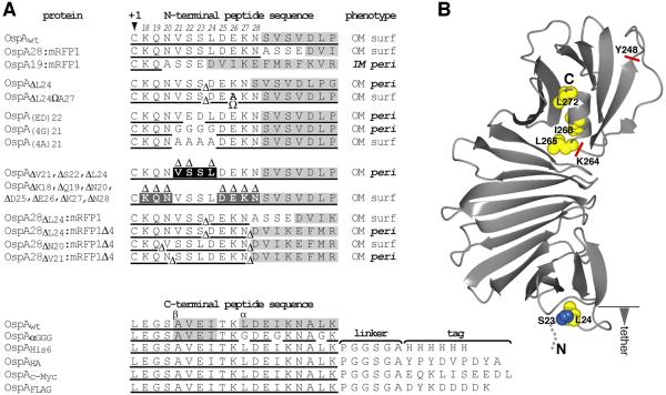
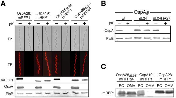
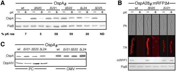
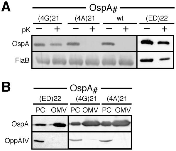
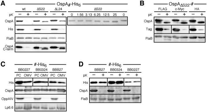
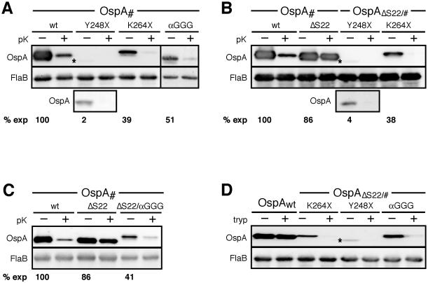
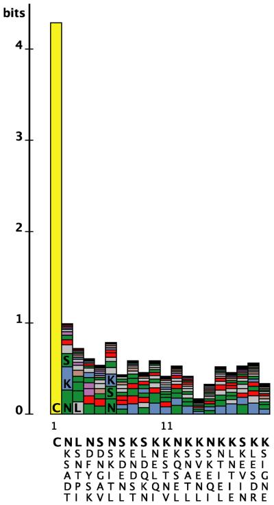
Borrelia burgdorferi surface lipoproteins are essential to the pathogenesis of Lyme borreliosis, but the mechanisms responsible for their localization are only beginning to emerge. We have previously demonstrated the critical nature of the amino-terminal 'tether' domain of the mature lipoprotein for sorting a fluorescent reporter to the Borrelia cell surface. Here, we show that individual deletion of four contiguous residues within the tether of major surface lipoprotein OspA results in its inefficient translocation across the Borrelia outer membrane. Intriguingly, C-terminal epitope tags of these N-terminal deletion mutants were selectively surface-exposed. Fold-destabilizing C-terminal point mutations and deletions did not block OspA secretion, but rather restored one of the otherwise periplasmic tether mutants to the bacterial surface. Together, these data indicate that disturbance of a confined tether feature leads to premature folding of OspA in the periplasm and thereby prevents secretion through the outer membrane. Furthermore, they suggest that OspA emerges tail-first on the bacterial surface, yet independent of a specific C-terminal targeting peptide sequence.


Figures

Figure 1
Figure 1. N- and C-terminal sequences of OspA lipoprotein mutants
(A) Deletions (Δ) and insertions (Ω) mutations are labelled with respect to the sequence of wild type (wt) OspA. Δ symbols within the sequence mark the deletion, and Δ symbols above the sequence indicate the deleted amino acid below. Please note that removal of either the Ser22 or Ser23 codon in the OspA tether yields identical peptides. Therefore, only the ΔSer22 mutant was generated. Gray shading indicates the structurally confined portion of the protein. Underlined sequence indicates the portion of the construct derived from wild type OspA. N-terminal mutant protein phenotypes are summarized by membrane (inner membrane, IM; outer membrane, OM), surface (surf) or periplasmic (peri) localization. (B) A ribbon representation of the OspA tertiary structure (PDB ID 1OSP, (Li et al., 1997) was generated using the CCP4 software for Macintosh (version 1.130.0; (Potterton et al., 2002). Mutated residues are highlighted as blue (Ser) or yellow (Leu or Ile) spheres representing the Cα and side chain atoms. Red lines indicate the sites of C-terminal truncations.
Figure 2
Figure 2. Role for OspA Leu24 in OM translocation
(A) Epifluorescence micrographs of B. burgdorferi expressing various red fluorescent protein fusions before and after treatment with proteinase K (pK). Ph, phase contrast; TR, Texas Red filter. Supporting Western immunoblots for mRFP1, using surface-exposed OspA and periplasmic FlaB as controls are shown below. (B) Proteinase K accessibility of OspA tether mutants compared to OspAwt. FlaB is used as a periplasmic, protease-resistant control. (C) Membrane fractionation immunoblots of OspAΔL24:mRFPΔ4 compared to surface-localized OspA28:mRFP1 and IM-localized OspA19:mRFP1 controls (Schulze and Zückert, 2006). OM, outer membrane vesicle fraction; PC, protoplasmic cylinder fraction (also containing intact cells; (Schulze and Zückert, 2006).
Figure 3
Figure 3. Localization of single-residue deletion OspA tether mutants
(A) Proteinase K (pK) accessibility assays for individual residue deletions from the OspA tether. Quantitative chemiluminescent immunoblot images were acquired using a Fuji LAS-4000 Luminescent Image Analyzer. The mean percentage of proteinase K resistance (% pK res) of OspA mutants was calculated from densitometry data from three independent in situ proteolysis assays with normalization to the FlaB signal. (B) Epifluorescence micrographs of B. burgdorferi B313 expressing various OspA tether-mRFPΔ4 fusions before and after treatment with proteinase K (pK). Ph, phase contrast; TR, Texas Red filter. Supporting Western immunoblots for mRFP1, using surface-exposed OspA and periplasmic FlaB as controls are shown below. (C) Membrane fractionation immunoblots of single-residue OspA deletions mutants compared to OspAwt. OppAIV served as IM control. OM, outer membrane vesicle fraction; PC, protoplasmic cylinder fraction (also containing intact cells; (Schulze and Zückert, 2006).
Figure 4
Figure 4. Localization of quadruple and double OspA tether substitution mutants
(A) Immunoblots of proteinase K (pK) accessibility assays for Val-Ser-Ser-Leu OspA tetrapeptide and Asp-Glu substitution OspA tether mutants. FlaB was used as a periplasmic, protease-resistant control. (B) Membrane fractionation immunoblots of the VSSL mutants compared to wild type OspA tether substitution mutants. OppAIV (Bono et al., 1998; Schulze and Zückert, 2006) served as IM control. OM, outer membrane vesicle fraction; PC, protoplasmic cylinder fraction (also containing intact cells; (Schulze and Zückert, 2006).
Figure 5
Figure 5. Localization of a C-terminal epitope tags in OspA tether deletion mutants
(A) Immunoblots of His6-tagged OspA subsurface mutants before and after proteinase K (pK) treatment. FlaB was used as a periplasmic, protease-resistant control. Note the slight downward shift of OspAΔS22-His6 and OspAΔL24-His6 anti-OspA-reacting bands and the concurrent loss of the His6 epitope upon in situ proteolytic treatment (left panel). To determine the amount necessary for cleavage of the His6 tag, OspAΔS22-His6 was subjected to shorter treatment with lower concentrations of protease (15 min vs. 1 h incubation with 1.56 to 50 μg/ml vs. the standard 200 μg/ml; right panel). (B) Immunoblots of OspA subsurface mutants with FLAG, c-Myc and HA C-terminal tags before and after proteinase K (pK) treatment. FlaB was used as a periplasmic, protease-resistant control. Note the tag accessibility phenotypes identical to the His6-tagged OspAΔS22 in panel A. (C) Membrane fractionation and (D) proteinase K (pK) accessibility immunoblots for C-terminally His6-tagged BB0227, BB0324, and BBB27 lipoproteins. OM, outer membrane vesicle fraction; PC, protoplasmic cylinder fraction (also containing intact cells; (Schulze and Zückert, 2006). Lipoproteins OppAIV (Bono et al., 1998; Schulze and Zückert, 2006) and Lp6.6 (Katona et al., 1992; Lahdenne et al., 1997) are included as IM and periplasmic OM controls, respectively. FlaB was used as periplasmic, protease-resistant control.
Figure 6
Figure 6. Localization of C-terminal fold-disrupting OspA mutants
(A) Immunoblots of proteinase K (pK) accessibility assays of C-terminal truncations and point mutants in otherwise wild type OspA. An asterisk labels the barely visible band for the OspAY248X mutant. To better demonstrate surface exposure, an immunoblot of a 5-fold concentrated sample is shown in the third row of the blot. Densitometry data of two independent in situ proteolysis assays yielded the mutants' mean expression level compared to OspAwt (% exp). (B) Proteinase K accessibility of C-terminal truncations in combination with an OspA tether ΔSer22 deletion. Surface accessibility of the OspAΔS22/Y248X mutant is shown as in panel A. (C) Protease accessibility of the C-terminal triple Gly substitution mutants. Protein stability data were obtained as in panel A. (D) Trypsin (tryp) susceptibility of surface-localized N- and C-terminal double mutants compared to wild type OspA. FlaB was used as periplasmic control in all panels.
Figure 7
Figure 7. Sequence complexity of B. burgdorferi lipoprotein tethers
A LogoBar (Perez-Bercoff et al., 2006) representation of the N-terminal sequence of known or predicted mature B. burgdorferi lipoproteins (Setubal et al., 2006) illustrates the complexity of the tether. The height of each column, measured in bits, is proportional to the lack of complexity at a given position. The columns are stacked from the bottom starting with the most frequently occurring residue at that position and continuing upward. Below each column are the six most frequently occurring residues at each position, in order of frequency from top (bold) to bottom. Colors represent residues with similar characteristics.

Similar articles

Cited by

References

    1. Babb K, McAlister JD, Miller JC, Stevenson B. Molecular characterization of Borrelia burgdorferi erp promoter/operator elements. J Bacteriol. 2004;186:2745–2756. - PMC - PubMed
    1. Barbour AG. Isolation and cultivation of Lyme disease spirochetes. Yale J Biol Med. 1984;57:521–525. - PMC - PubMed
    1. Barbour AG, Hayes SF, Heiland RA, Schrumpf ME, Tessier SL. A Borrelia-specific monoclonal antibody binds to a flagellar epitope. Infect Immun. 1986;52:549–554. - PMC - PubMed
    1. Barbour AG, Tessier SL, Hayes SF. Variation in a major surface protein of Lyme disease spirochetes. Infect Immun. 1984;45:94–100. - PMC - PubMed
    1. Barbour AG, Tessier SL, Todd WJ. Lyme disease spirochetes and ixodid tick spirochetes share a common surface antigenic determinant defined by a monoclonal antibody. Infect Immun. 1983;41:795–804. - PMC - PubMed

Publication types

MeSH terms

LinkOut - more resources