

Crystal structure of Helicobacter pylori MinE, a cell division topological specificity factor
- PMID: 20398219
- PMCID: PMC2883074
- DOI: 10.1111/j.1365-2958.2010.07160.x
Crystal structure of Helicobacter pylori MinE, a cell division topological specificity factor
Abstract
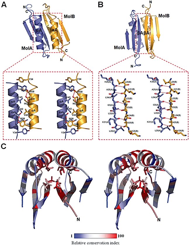
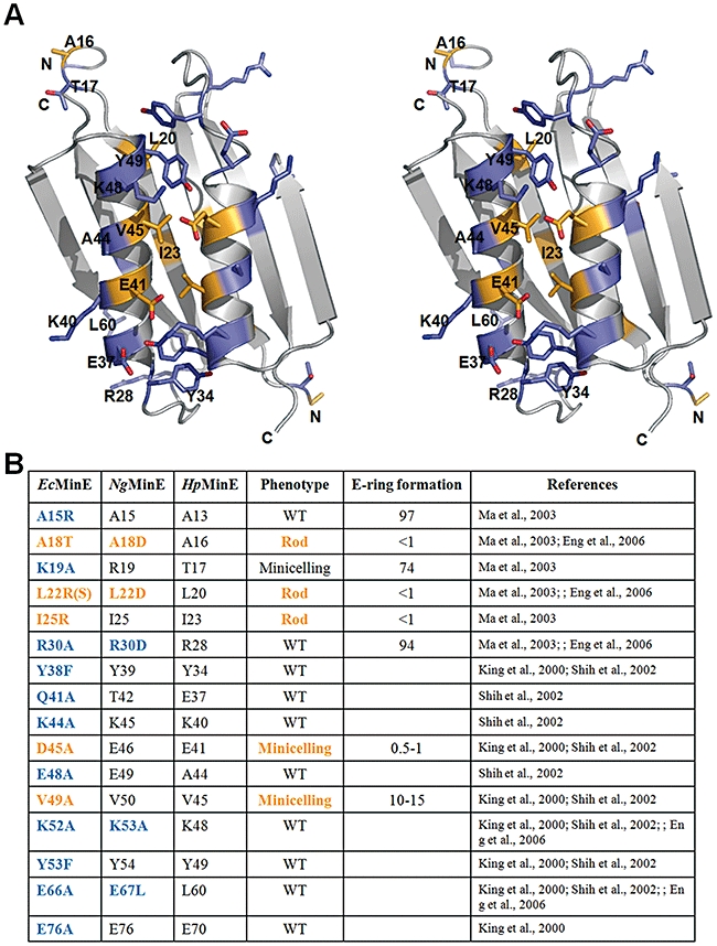
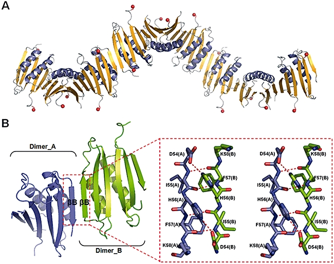
In Gram-negative bacteria, proper placement of the FtsZ ring, mediated by nucleoid occlusion and the activities of the dynamic oscillating Min proteins MinC, MinD and MinE, is required for correct positioning of the cell division septum. MinE is a topological specificity factor that counters the activity of MinCD division inhibitor at the mid-cell division site. Its structure consists of an anti-MinCD domain and a topology specificity domain (TSD). Previous NMR analysis of truncated Escherichia coli MinE showed that the TSD domain contains a long alpha-helix and two anti-parallel beta-strands, which mediate formation of a homodimeric alpha/beta structure. Here we report the crystal structure of full-length Helicobacter pylori MinE and redefine its TSD based on that structure. The N-terminal region of the TSD (residues 19-26), previously defined as part of the anti-MinCD domain, forms a beta-strand (betaA) and participates in TSD folding. In addition, H. pylori MinE forms a dimer through the interaction of anti-parallel betaA-strands. Moreover, we observed serial dimer-dimer interactions within the crystal packing, resulting in the formation of a multimeric structure. We therefore redefine the functional domain of MinE and propose that a multimeric filamentous structure is formed through anti-parallel beta-strand interactions.
Figures

Similar articles
-
Conformation of the cell division regulator MinE: evidence for interactions between the topological specificity and anti-MinCD domains.Biochemistry. 2006 Apr 11;45(14):4593-601. doi: 10.1021/bi060022j. Biochemistry. 2006. PMID: 16584194
-
Crystallization and preliminary X-ray crystallographic analysis of MinE, the cell-division topological specificity factor from Helicobacter pylori.Acta Crystallogr Sect F Struct Biol Cryst Commun. 2010 May 1;66(Pt 5):527-9. doi: 10.1107/S1744309110009784. Epub 2010 Apr 29. Acta Crystallogr Sect F Struct Biol Cryst Commun. 2010. PMID: 20445251 Free PMC article.
-
Structural basis for the topological specificity function of MinE.Nat Struct Biol. 2000 Nov;7(11):1013-7. doi: 10.1038/80917. Nat Struct Biol. 2000. PMID: 11062554
-
The dimerization and topological specificity functions of MinE reside in a structurally autonomous C-terminal domain.Mol Microbiol. 1999 Feb;31(4):1161-9. doi: 10.1046/j.1365-2958.1999.01256.x. Mol Microbiol. 1999. PMID: 10096083
-
Sticky socks: Helicobacter pylori VacA takes shape.Trends Microbiol. 2008 Mar;16(3):89-92. doi: 10.1016/j.tim.2008.01.001. Epub 2008 Feb 14. Trends Microbiol. 2008. PMID: 18280164 Review.
Cited by
-
The Min oscillator uses MinD-dependent conformational changes in MinE to spatially regulate cytokinesis.Cell. 2011 Aug 5;146(3):396-407. doi: 10.1016/j.cell.2011.06.042. Cell. 2011. PMID: 21816275 Free PMC article.
-
Regulation of symmetric bacterial cell division by MinE: What is the role of conformational dynamics?Commun Integr Biol. 2011 Jan;4(1):101-3. doi: 10.4161/cib.4.1.14162. Commun Integr Biol. 2011. PMID: 21509194 Free PMC article.
-
Acid-regulated gene expression of Helicobacter pylori: Insight into acid protection and gastric colonization.Helicobacter. 2018 Jun;23(3):e12490. doi: 10.1111/hel.12490. Epub 2018 Apr 25. Helicobacter. 2018. PMID: 29696729 Free PMC article.
-
Molecular Interactions of the Min Protein System Reproduce Spatiotemporal Patterning in Growing and Dividing Escherichia coli Cells.PLoS One. 2015 May 27;10(5):e0128148. doi: 10.1371/journal.pone.0128148. eCollection 2015. PLoS One. 2015. PMID: 26018614 Free PMC article.
-
Appropriation of the MinD protein-interaction motif by the dimeric interface of the bacterial cell division regulator MinE.Proc Natl Acad Sci U S A. 2010 Oct 26;107(43):18416-21. doi: 10.1073/pnas.1007141107. Epub 2010 Oct 11. Proc Natl Acad Sci U S A. 2010. PMID: 20937912 Free PMC article.
References
-
- de Boer PA, Crossley RE, Rothfield LI. A division inhibitor and a topological specificity factor coded for by the minicell locus determine proper placement of the division septum in E. coli. Cell. 1989;56:641–649. - PubMed
-
- Brunger AT, Adams PD, Clore GM, DeLano WL, Gros P, GrosseKunstleve RW, et al. Crystallography & NMR system: a new software suite for macromolecular structure determination. Acta Crystallogr D Biol Crystallogr. 1998;54:905–921. - PubMed
-
- DeLano WL. The PyMOL Molecular Graphics System, 0.99 Ed. Palo Alto, CA: DeLano Scientific; 2002.
Publication types
MeSH terms
Substances
LinkOut - more resources
Full Text Sources
