

Modulation of SOCS protein expression influences the interferon responsiveness of human melanoma cells
- PMID: 20398276
- PMCID: PMC2858748
- DOI: 10.1186/1471-2407-10-142
Modulation of SOCS protein expression influences the interferon responsiveness of human melanoma cells
Abstract
Background: Endogenously produced interferons can regulate the growth of melanoma cells and are administered exogenously as therapeutic agents to patients with advanced cancer. We investigated the role of negative regulators of interferon signaling known as suppressors of cytokine signaling (SOCS) in mediating interferon-resistance in human melanoma cells.
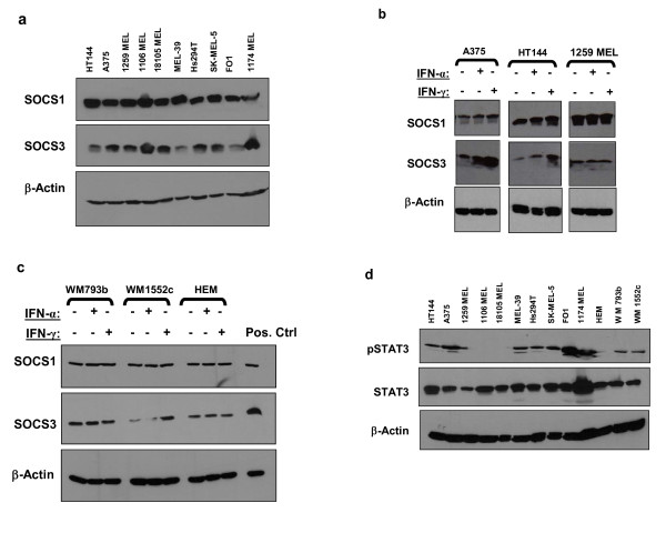
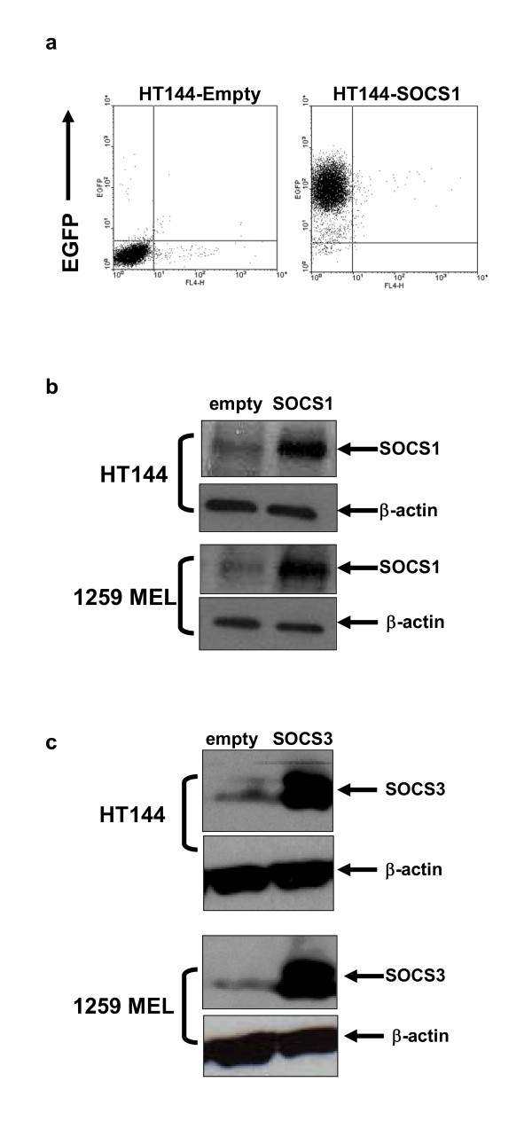
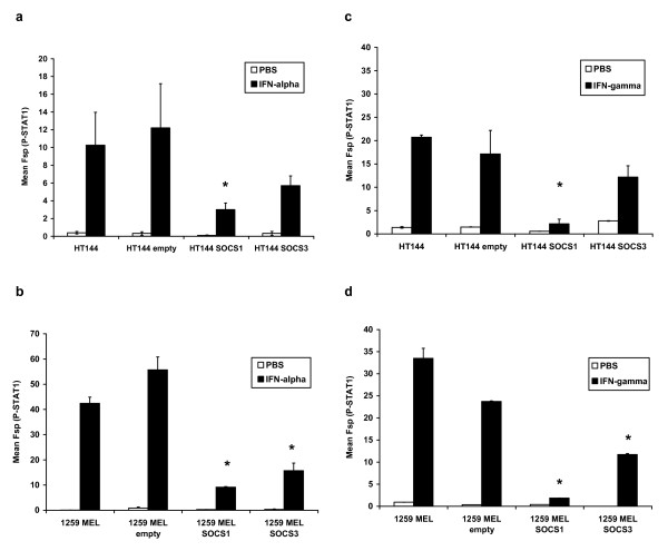
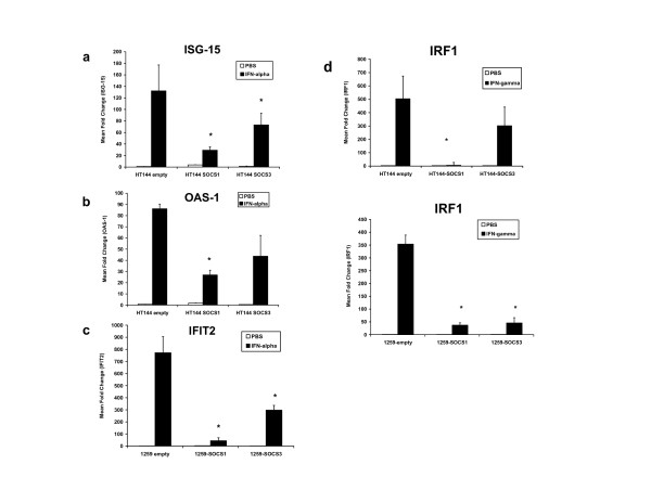
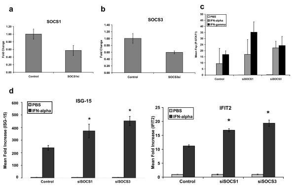
Methods: Basal and interferon-alpha (IFN-alpha) or interferon-gamma (IFN-gamma)-induced expression of SOCS1 and SOCS3 proteins was evaluated by immunoblot analysis in a panel of n = 10 metastatic human melanoma cell lines, in human embryonic melanocytes (HEM), and radial or vertical growth phase melanoma cells. Over-expression of SOCS1 and SOCS3 proteins in melanoma cells was achieved using the PINCO retroviral vector, while siRNA were used to inhibit SOCS1 and SOCS3 expression. Tyr701-phosphorylated STAT1 (P-STAT1) was measured by intracellular flow cytometry and IFN-stimulated gene expression was measured by Real Time PCR.
Results: SOCS1 and SOCS3 proteins were expressed at basal levels in melanocytes and in all melanoma cell lines examined. Expression of the SOCS1 and SOCS3 proteins was also enhanced following stimulation of a subset of cell lines with IFN-alpha or IFN-gamma. Over-expression of SOCS proteins in melanoma cell lines led to significant inhibition of Tyr701-phosphorylated STAT1 (P-STAT1) and gene expression following stimulation with IFN-alpha (IFIT2, OAS-1, ISG-15) or IFN-gamma (IRF1). Conversely, siRNA inhibition of SOCS1 and SOCS3 expression in melanoma cells enhanced their responsiveness to interferon stimulation.
Conclusions: These data demonstrate that SOCS proteins are expressed in human melanoma cell lines and their modulation can influence the responsiveness of melanoma cells to IFN-alpha and IFN-gamma.
Figures
Similar articles
-
Interferon-gamma, but not interferon-alpha, induces SOCS 3 expression in human melanoma cell lines.Melanoma Res. 2005 Dec;15(6):481-8. doi: 10.1097/00008390-200512000-00001. Melanoma Res. 2005. PMID: 16314732
-
IFN-alpha-induced signal transduction, gene expression, and antitumor activity of immune effector cells are negatively regulated by suppressor of cytokine signaling proteins.J Immunol. 2007 Apr 15;178(8):4832-45. doi: 10.4049/jimmunol.178.8.4832. J Immunol. 2007. PMID: 17404264
-
The suppressor of cytokine signaling (SOCS) 1 and SOCS3 but not SOCS2 proteins inhibit interferon-mediated antiviral and antiproliferative activities.J Biol Chem. 1998 Dec 25;273(52):35056-62. doi: 10.1074/jbc.273.52.35056. J Biol Chem. 1998. PMID: 9857039
-
SOCS, Intrinsic Virulence Factors, and Treatment of COVID-19.Front Immunol. 2020 Oct 23;11:582102. doi: 10.3389/fimmu.2020.582102. eCollection 2020. Front Immunol. 2020. PMID: 33193390 Free PMC article. Review.
-
Complexity of Interferon-γ Interactions with HSV-1.Front Immunol. 2014 Feb 6;5:15. doi: 10.3389/fimmu.2014.00015. eCollection 2014. Front Immunol. 2014. PMID: 24567732 Free PMC article. Review.
Cited by
-
SOCS, inflammation, and cancer.JAKSTAT. 2013 Jul 1;2(3):e24053. doi: 10.4161/jkst.24053. Epub 2013 Aug 15. JAKSTAT. 2013. PMID: 24069550 Free PMC article. Review.
-
Tumor-infiltrating T lymphocytes improve clinical outcome of therapy-resistant neuroblastoma.Oncoimmunology. 2015 Apr 2;4(9):e1019981. doi: 10.1080/2162402X.2015.1019981. eCollection 2015 Sep. Oncoimmunology. 2015. PMID: 26405592 Free PMC article.
-
Cigarette smoke-induced lung inflammation in COPD mediated via LTB4/BLT1/SOCS1 pathway.Int J Chron Obstruct Pulmon Dis. 2015 Dec 22;11:31-41. doi: 10.2147/COPD.S96412. eCollection 2016. Int J Chron Obstruct Pulmon Dis. 2015. PMID: 26730186 Free PMC article.
-
IRF1 and NF-kB restore MHC class I-restricted tumor antigen processing and presentation to cytotoxic T cells in aggressive neuroblastoma.PLoS One. 2012;7(10):e46928. doi: 10.1371/journal.pone.0046928. Epub 2012 Oct 5. PLoS One. 2012. PMID: 23071666 Free PMC article.
-
SOCS1 and SOCS3 as key checkpoint molecules in the immune responses associated to skin inflammation and malignant transformation.Front Immunol. 2024 Jun 21;15:1393799. doi: 10.3389/fimmu.2024.1393799. eCollection 2024. Front Immunol. 2024. PMID: 38975347 Free PMC article. Review.
References
-
- Kirkwood JM, Ibrahim JG, Sondak VK, Richards J, Flaherty LE, Ernstoff MS, Smith TJ, Rao U, Steele M, Blum RH. High- and low-dose interferon alfa-2b in high-risk melanoma: first analysis of intergroup trial E1690/S9111/C9190. J Clin Oncol. 2000;18(12):2444–2458. - PubMed
Publication types
MeSH terms
Substances
Grants and funding
LinkOut - more resources
Full Text Sources
Other Literature Sources
Medical
Research Materials
Miscellaneous
