Review
. 2010 Apr 19:12:e12.
doi: 10.1017/S1462399410001456.

Mitochondrial quality control and neurological disease: an emerging connection

Affiliations
Free PMC article
Review

Mitochondrial quality control and neurological disease: an emerging connection

Inês Pimenta de Castro et al. Expert Rev Mol Med. .
Free PMC article

Abstract

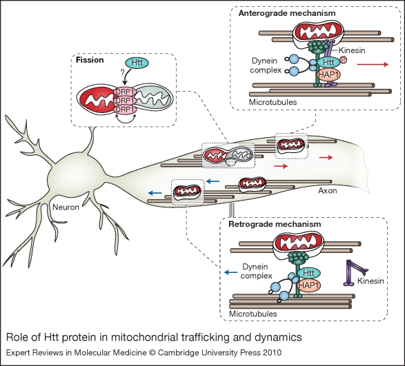
The human brain is a highly complex organ with remarkable energy demands. Although it represents only 2% of the total body weight, it accounts for 20% of all oxygen consumption, reflecting its high rate of metabolic activity. Mitochondria have a crucial role in the supply of energy to the brain. Consequently, their deterioration can have important detrimental consequences on the function and plasticity of neurons, and is thought to have a pivotal role in ageing and in the pathogenesis of several neurological disorders. Owing to their inherent physiological functions, mitochondria are subjected to particularly high levels of stress and have evolved specific molecular quality-control mechanisms to maintain the mitochondrial components. Here, we review some of the most recent advances in the understanding of mitochondrial stress-control pathways, with a particular focus on how defects in such pathways might contribute to neurodegenerative disease.


Figures

Figure 1
Figure 1
Core quality-control pathways in mitochondria. Low levels of damaged proteins in mitochondria are cleared at the molecular level by intraorganellar proteases and chaperones such as OMI/HTRA2 and TRAP1 (top left). Enhanced levels of damage probably overwhelm the capacity of the molecular quality-control machinery (middle left), leading to the proposed segregation of damaged mitochondrial components by the fusion/fission machinery. This enables the physical separation of healthy (green) and damaged (orange) daughter mitochondria. Damaged mitochondria are then recycled using the cellular autophagy pathyways. If the levels of damage exceed the capacity of both molecular and organellar quality-control pathways (top right), mitochondria can rupture, leading to the release of apoptosis-promoting factors and cell death.
Figure 2
Figure 2
Molecular determinants of mitochondrial quality-control networks. Mitochondria are dynamic organelles that are continuously remodelled by fusion and fission events. Fusion requires the joining of both the inner and outer mitochondrial membranes of two mitochondria. Mitofusin 1 (MFN1) and mitofusin 2 (MFN2) carry out the initial joining of the outer membrane fusion, whereas OPA1 mediates the fusion of the inner membrane. Mitochondrial fission is mediated by DRP1 and FIS1; DRP1 oligomerizes into 8–12 subunit chains and is recruited to mitochondria via the adaptor FIS1. This recruitment allows the full oligomerisation of DRP1 into spiral chains that wrap around the mitochondria and, by constriction, causes mitochondrial fission. In this figure, fission is shown as a means to selectively eliminate damaged mitochondrial components by packing them in one of the fission products, and it has been proposed to involve the asymmetrical segregation of OPA1 (Ref. 5). In this context, fission produces metabolically different daughter units that may either maintain (red) or lose (grey) an intact membrane potential. Depolarised mitochondria are targeted to degradation by autophagy (mitophagy) through a mechanism that is still poorly defined. Here, we illustrate two possibilities: (1) the presence of proteins associated with mitochondrial membranes, such as the mammalian BNIP3L/NIX (Ref. 109, 110), and the yeast proteins Uth1p (Ref. 111) and Aup1 (Ref. 112), which are involved in the direct entrapment of mitochondria by autophagosomes; (2) the activity of E3 ligases, such as LISTERIN (Ref. 113), MULAN (Ref. 114) and Parkin (Refs 28, 115), which might regulate the conjugation of monoubiquitin (Ub) or polyubiquitin chains to an exposed mitochondrial protein, thereby targeting the organelle to autophagic degradation. p62 and NBR1 are autophagic receptors that bind both Ub and ATG8/LC3 on the phagophore (Ref. 116), which might constitute the mechanistic link between mitochondrial ubiquitylation and mitophagy. The mTOR signalling pathway acts as a major positive modulator of mitochondrial metabolism and biogenesis. We propose that the inhibition of mTORC1 upon mitochondrial damage caused by loss of ATP and consequential activation of AMPK, might be crucial to ensure an enhancement of the autophagic degradation of defective mitochondria. Abbreviations: AMPK, AMP-activated protein kinase; Aup1p, yeast mitochondrial protein phosphatase homologue; BNIP3L/NIX, BCL2/adenovirus E1B 19 kDa protein-interacting protein 3-like; LC3, light chain 3 protein; LKB1, serine/threonine-protein kinase 11; mTOR, mammalian target of rapamycin; mTORC1, mTOR complex 1; NBR1, next to BRCA1 gene 1 protein; OM, outer membrane; OPA1, optic atrophy 1; Uth1p, yeast outer mitochondrial membrane protein.
Figure 3
Figure 3
Role of the major PD-associated genes in mitochondrial dysfunction. Many genes associated with Parkinson disease (PD) implicate a role for mitochondria in the pathogenesis of the disease. The serine protease HTRA2–OMI is synthesised as an inactive precursor containing a mitochondrial-targeting sequence (MTS). In response to the activation of the MEKK3–p38 stress-kinase-signalling pathway, HTRA2/OMI is phosphorylated by p38 in a PINK1-dependent manner and imported into the intermembrane space (IMS), where it has been reported to bind to the Bcl-2-family-related protein Hax-1 (Ref. 117). Despite the fact that this interaction is controversial (Ref. 118), it has been suggested to promote the proteolytic processing of HTRA2/OMI by the mitochondrial protease PARL. Active HTRA2/OMI is thought to be involved in the degradation of misfolded proteins present in the IMS (Ref. 119) and to prevent the oligomerisation of the activated form of BAX on the outer membrane (OM), thus avoiding apoptosis (Ref. 117). Under oxidative stress conditions, PINK1 also interacts with the mitochondrial molecular chaperone TRAP1. Once phosphorylated, TRAP1 inhibits oxidative-stress-induced cytochrome c (Cyt C) release, prevents misfolding and promotes the correct assembly of mitochondrial proteins. DJ-1 is a cytosolic oxidative-stress-regulated chaperone, which redistributes from the cytosol to the mitochondria upon oxidation of specific cysteine residues. In the cytoplasm, DJ-1 prevents the aggregation and toxicity of α-synuclein (α-syn); in mitochondria, it has been suggested to protect respiratory complex I (cI) from oxidative-stress-mediated inactivation (Ref. 23). Another protein involved in the mitochondrial stress response is Parkin, a cytosolic E3-ubiquitin ligase, which is selectively recruited to uncoupled or dysfunctional mitochondria, targeting them for autophagy (Ref. 28). A model is illustrated whereby PINK1, acting upstream of Parkin, might regulate mitochondrial function by sensing mitochondrial damage, recruiting Parkin and inducing mitophagy. Abbreviations: ADP, adenosine diphosphate; ATP, adenosine-5-triphosphate; cII, complex II; cIII, complex III; cIV, complex IV, cV, complex V; HAX-1, HCLS1-associated protein X-1; HTRA2, high-temperature requirement A2; IM, mitochondrial inner membrane; MEKK3, mitogen-activated protein kinase kinase kinase 3; PINK1, PTEN-induced putative kinase 1; PD, Parkinson disease; PDZ domain, post-synaptic density protein (PSD95), Drosophila discs large tumour suppressor (DlgA) and zonula occludens-1 protein (zo-1) domain; TRAP1, TNF-receptor associated protein; Δψm, mitochondrial membrane potential.
Figure 4
Figure 4
Mitochondrial dysfunction in AD. Degenerating neurons in brain areas affected by Alzheimer disease (AD) (e.g. hippocampus and cerebral cortex) present typical intracellular neurofibrillary tangles (NFTs) and extracellular accumulation of β-amyloid plaques. Aβ is produced from the metabolism of amyloid precursor protein (APP) through sequential cleavage by β- and γ-secretases, and it is prone to form toxic oligomeric structures both inside and outside the cell. APP and Aβ can affect mitochondrial function by different mechanisms. APP is targeted to mitochondria, where it forms complexes with the translocases of the outer and inner membranes (TOM and TIM), which drive the import of mitochondrial proteins in cooperation with molecular chaperones. However, the presence of an acidic domain within APP might be responsible for preventing its translocation into mitochondria. As a consequence, the import of mitochondrial proteins, such as respiratory chain subunits, is reduced, and this event is associated with increased free radical generation and reduced activity of the electron-transport chain. Other aspects of amyloid metabolism involve mitochondria: intramitochondrial Aβ has been shown to interact with amyloid-β-binding alcohol dehydrogenase (ABAD) and to produce reactive oxygen species (ROS). Aβ also interacts with cytochrome c oxidase, thus decreasing the activity of complex IV (cIV). Furthermore, presequence peptidase PreP and HTRA2/OMI serine protease have been shown to degrade Aβ oligomers, thus providing a mechanism to detoxify this metabolite. Intramitochondrial Aβ directly interacts with cyclophilin D (CypD), a component of the mitochondrial permeability transition pore (mPTP), which is located in the mitochondrial matrix. This interaction makes the channel more sensitive to Ca2+ and stimulates mPTP opening, thus raising the permeability of the mitochondrial inner membrane (IM) and eventually disrupting the mitochondrial outer membrane (OM). As a result, deregulation of the mPTP opening determines a functional disorder that triggers cell death. Abbreviations: AD, Alzheimer disease; ATP, adenosine-5′-triphosphate; cI, complex I; cII, complex II; cIII, complex III; cV, complex V; HTRA2, HtrA2 serine peptidase; Δψm, mitochondrial membrane potential.
Figure 5
Figure 5
Role of Htt protein in mitochondrial trafficking and dynamics. Mitochondrial movement in neurons is highly diverse and complex. Normal Htt protein regulates anterograde (away from the cell body) and retrograde (towards the cell body) transport of mitochondria by interacting with several trafficking mediators. Htt stimulates trafficking by binding to HAP1, which in turn, leads to interaction with the motor proteins dynein–dynactin and kinesin. Phosphorylation of Htt acts as a molecular switch for anterograde versus retrograde mitochondrial transport. When Htt is phosphorylated, kinesin-1 is recruited and promotes anterograde transport; conversely, when Htt is unphosphorylated, kinesin-1 detaches from the motor complex and induces a switch to retrograde transport (Ref. 95). In addition to migration and movement, mitochondria undergo cycles of fusion and fission. The key mitochondrial fission regulator is dynamin-related protein 1 (DRP1). Similarly to dynamin, DRP1 seems to act as a mechano-enzyme to constrict and divide mitochondria. Given that Htt interacts with dynamin, one can speculate that Htt might regulate fission by interacting with DRP1. Abbreviations: HAP1, Htt-associated protein 1; Htt, Huntingtin protein; P, phosphate.

Similar articles

Cited by

References

    1. Lane N. Power, Sex, Suicide: mitochondria and the meaning of life. Oxford University Press; Oxford: 2005.
    1. Chan D.C.. Mitochondria: dynamic organelles in disease, aging, and development. Cell. 2006;125:1241–1252. - PubMed
    1. Taylor S.W., Fahy E.. Ghosh S.S.. Global organellar proteomics. Trends in Biotechnology. 2003;21:82–88. and . - PubMed
    1. Wang Y.. Bogenhagen D.F.. Human mitochondrial DNA nucleoids are linked to protein folding machinery and metabolic enzymes at the mitochondrial inner membrane. Journal of Biological Chemistry. 2006;281:25791–25802. and . - PubMed
    1. Twig G., Hyde B.. Shirihai O.S.. Mitochondrial fusion, fission and autophagy as a quality control axis: the bioenergetic view. Biochimica et Biophysica Acta. 2008;1777:1092–1097. and . - PMC - PubMed

Further reading, resources and contacts

Books
    1. Lane N. Power, Sex, Suicide: Mitochondria and the Meaning of Life. OUP; Oxford: 2005.
    2. A provocative book full of interesting concepts, including links between ageing and neurodegenerative disease.

    1. Gibson G.E., Ratan R.R.. Flint Beal M.. Mitochondria and Oxidative Stress in Neurodegenerative Disorders. Annals of the New York Academy of Sciences 2008 and . eds ( - PubMed
    2. A combination of basic and clinical research to give the reader the most current information on aspects of mitochondrial function linked to age-related neurodegenerative diseases and their treatment.

Websites
    1. http://www.alz.org http://www.alz.org
    2. A database of Drosophila melanogaster nuclear genes encoding mitochondrial proteins can be found at:

    1. http://www2.ba.itb.cnr.it/MitoDrome http://www2.ba.itb.cnr.it/MitoDrome
    2. The Whitehead Institute Video Gallery has lectures from researchers at the Whitehead Institute. It includes several interesting lectures by Susan Lindquist on protein misfolding and neurodegenerative diseases as well as David Sabatini on growth control pathways:

    1. www.wi.mit.edu/news/video_gallery www.wi.mit.edu/news/video_gallery
    2. The Encyclopedia of Neuroscience explores a wide variety of topics related to different areas of neuroscience. It is a very useful tool, well written and easily accessible:

    1. www.sciencedirect.com/science/referenceworks/9780080450469 www.sciencedirect.com/science/referenceworks/9780080450469
    2. The Allen Mouse Brain Atlas is an interactive, genome-wide image database of gene expression. It also includes information regarding the cortex of the human brain:

    1. www.mouse.brain-map.org www.mouse.brain-map.org

Publication types

MeSH terms

Substances