. 2010 Jul 1;343(1-2):40-50.
doi: 10.1016/j.ydbio.2010.04.005. Epub 2010 Apr 14.

Differential sensitivity of epidermal cell subpopulations to beta-catenin-induced ectopic hair follicle formation

Affiliations

Differential sensitivity of epidermal cell subpopulations to beta-catenin-induced ectopic hair follicle formation

Christopher M Baker et al. Dev Biol. .

Abstract

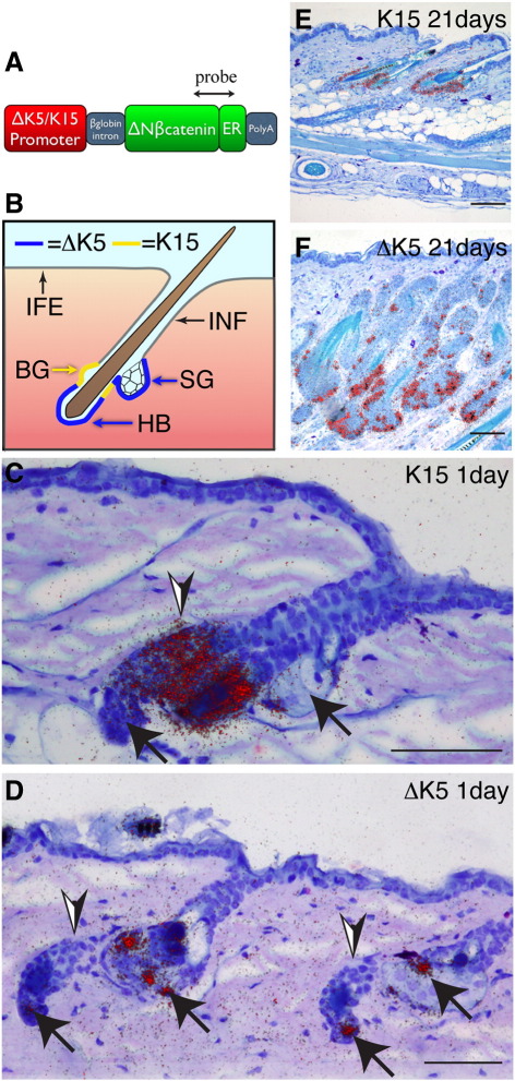
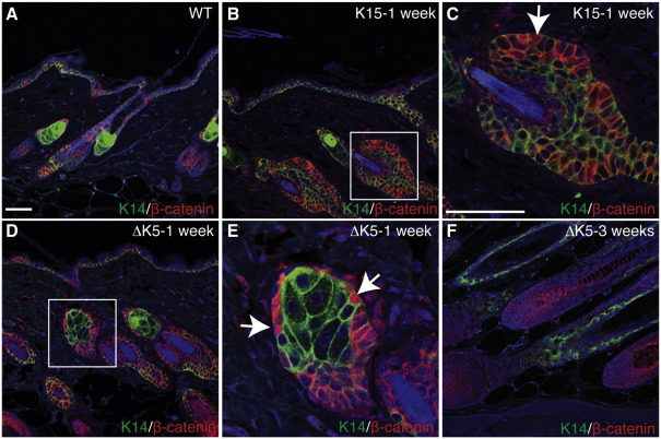
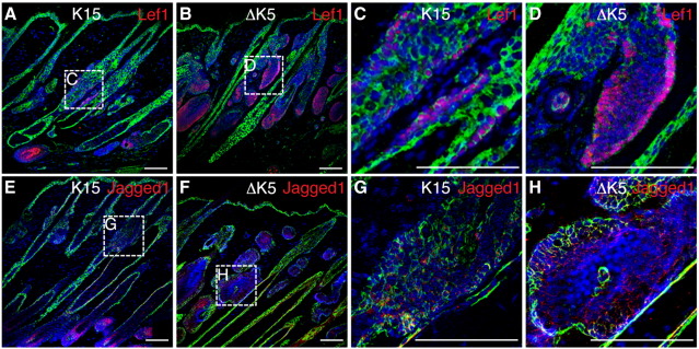
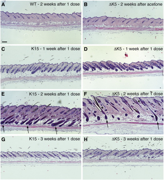
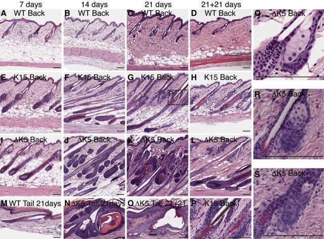
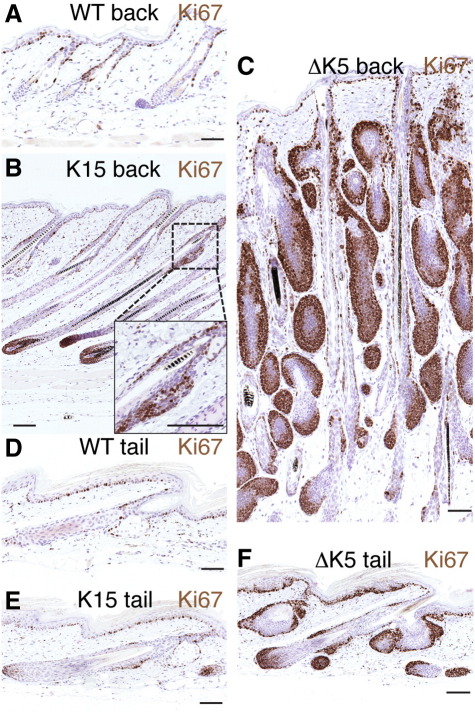
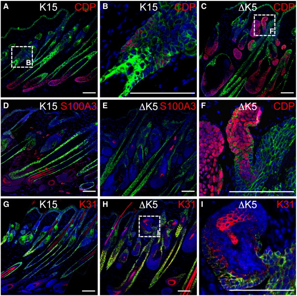
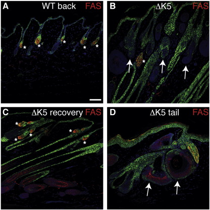
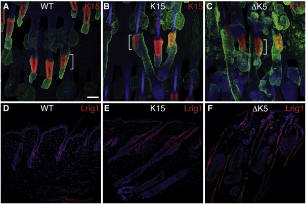
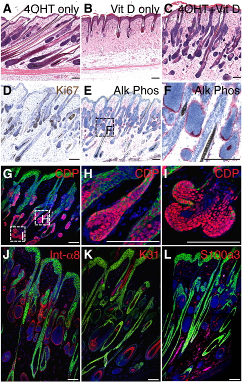
Wnt signalling is required for hair follicle development and for the growth phase (anagen) of postnatal follicles. When the pathway is activated at high levels in adult mouse epidermis, ectopic follicles form from existing follicles, interfollicular epidermis (IFE) and sebaceous glands, revealing a remarkable ability of the tissue to be reprogrammed. To compare the competence of different epidermal cell populations to form ectopic follicles, we expressed a 4-hydroxy-tamoxifen (4OHT) inducible, stabilised beta-catenin transgene (DeltaNbeta-cateninER) under the control of two different promoters. We targeted the reservoir of stem cells in the hair follicle bulge via the keratin 15 (K15) promoter and targeted the sebaceous glands and base of the follicle (bulb) with a truncated K5 promoter (DeltaK5). No ectopic follicles formed in the IFE in either model, establishing the autonomy of the IFE stem cell compartment in undamaged epidermis. Activation of beta-catenin in the bulge stimulated proliferation and bulge expansion. Existing hair follicles entered anagen, but no ectopic follicles formed. DeltaK5DeltaNbeta-cateninER expressing hair follicles also entered anagen on 4OHT treatment. In addition, a subpopulation of cells at the base of the sebaceous gland readily formed ectopic follicles, resulting in complete and reversible conversion of sebaceous glands into hair follicles. Combined activation of beta-catenin and the vitamin D receptor enhanced differentiation of sebaceous gland-derived hair follicles and stimulated ectopic follicle formation in the hair follicle bulb, but not in the bulge. Our results suggest that the bulge and sebaceous gland are, respectively, non-permissive and permissive niches for Wnt induced hair follicle differentiation.


Figures

Fig. 1
Fig. 1
Sites of transgene expression. (A) Schematic of the ΔK5 and K15ΔNβ-cateninER constructs. Double headed arrow indicates region detected with the in situ hybridisation probe. (B) Schematic showing sites of promoter activity. The ΔK5 promoter targets the hair follicle bulb (HB) and sebaceous glands (SG) but not the bulge (BG). The K15 promoter targets the hair follicle bulge. Neither promoter is active in the interfollicular epidermis (IFE) or infundibulum (INF). (C–F) In situ hybridisation of back skin sections of K15ΔNβ-cateninER (K15) and ∆K5ΔNβ-cateninER (ΔK5) transgenic mouse lines treated with 4OHT for (C, D) 1 day or (E, F) 21 days. Arrowheads indicate position of bulge; arrows mark hair bulb and sebaceous glands. Silver grains in darkfield images are false coloured red, superimposed on haemotoxylin staining. Scale bars: 50 µm.
Fig. 2
Fig. 2
Location of cells with nuclear β-catenin. Sections of wild type telogen back skin (A) or transgenic back skin that had received repeated 4OHT applications for the number of weeks shown (B–F) were labeled with antibodies to keratin 14 (green) and β-catenin (red) and counterstained with DAPI (blue). Boxed regions in B, D are shown at higher magnification in C, E, respectively. Arrows indicate β-catenin positive nuclei. (F) Nuclear β-catenin in cells at the base of an anagen follicle. Scale bar: 100 µm.
Fig. 3
Fig. 3
Expression of β-catenin target genes Lef1 and Jagged1. Immunofluorescence labeling of sections of K15ΔNβ-cateninER (K15) and ∆K5ΔNβ-cateninER (ΔK5) back skin treated with 4OHT for 21 days. Antibodies detected (A–D) Lef1 (red), (E–H) Jagged1 (red). Sections were also labeled with anti-keratin 14 (green) and counterstained with DAPI (blue) to visualise nuclei. Boxed regions in A, B, E, F are shown at higher magnification in C, D, G, H, respectively. Scale bars: 50 µm.
Fig. 4
Fig. 4
Effects of a single dose of 4OHT. Haematoxylin and eosin stained sections of back skin from telogen mice that had been treated once with acetone (B) or 4OHT (A, C–H) and collected 1–3 weeks later. WT: wild type. Arrows show anagen follicles. Asterisks show ectopic follicles. Scale bar: 100 µm.
Fig. 5
Fig. 5
Effects of repeated doses of 4OHT. Haematoxylin and eosin stained sections of wild type (WT), K15ΔNβ-cateninER (K15) and ∆K5ΔNβ-cateninER (ΔK5) transgenic skin treated with 4OHT for 7 days (A, E, I, Q, R), 14 days (B, F, J, S), 21 days (C, G, K, M, N, P), or 21 days + 21 days recovery (D, H, L, O). Note cysts in (N, O) and conversion of sebaceous glands into ectopic hair follicles in (Q–S). Scale bars: 50 µm.
Fig. 6
Fig. 6
Activation of β-catenin stimulates proliferation in transgene positive epidermal cells. Immunohistochemical labeling with anti-Ki67 (brown) of back and tail skin from wild type (WT) (A, D), K15ΔNβ-cateninER (K15) (B, E) and ∆K5ΔNβ-cateninER (ΔK5) (C, F) transgenic mice following treatment with 4OHT for 21 days. Scale bars: 50 µm.
Fig. 7
Fig. 7
Expression of hair follicle differentiation markers. Immunofluorescence staining of K15ΔNβ-cateninER (K15) and ∆K5ΔNβ-cateninER (ΔK5) transgenic back skin following 21 days of 4OHT treatment. Sections were labeled with antibodies to (A–C, F) CDP (red), (D, E) S100A3 (red) or (G–I) K31 (red), and keratin 14 (green), with DAPI nuclear counterstain (blue). Scale bars: 50 µm.
Fig. 8
Fig. 8
Sebaceous gland differentiation. Immunofluorescence staining of wild type (WT) and ∆K5ΔNβ-cateninER (ΔK5) transgenic back (A–C) and tail (D) skin following 21 days of 4OHT treatment. Skin in (C) was left untreated for a further 21 days. Sections were labeled with antibodies fatty acid synthase (red) and keratin 14 (green), with DAPI nuclear counterstain (blue). Asterisks indicate fatty acid synthase positive regions. Arrows in (B) indicate ectopic HF arising from SG. Arrows in (D) indicate cysts. Scale bar: 50 µm.
Fig. 9
Fig. 9
Expression of stem cell markers K15 and Lrig1. Immunofluorescence staining of wild type (WT), K15ΔNβ-cateninER (K15) and ∆K5ΔNβ-cateninER (ΔK5) transgenic skin following 21 days of 4OHT treatment. (A–C) Tail skin whole mounts labeled for K15 (red) and K14 (green) with DAPI nuclear counterstain (blue). Brackets indicate position of bulge in existing follicles. (D–F) Back skin sections labeled with anti-Lrig1 (red) with DAPI nuclear counterstain (blue). Scale bar: 50 µm.
Fig. 10
Fig. 10
Effects of combined activation of β-catenin and vitamin D receptor. Back skin of ∆K5ΔNβ-cateninER transgenic mice was treated with (A) 4OHT alone, (B) 1,25-dihydroxyvitamin D3 analogue alone (Vit D) or (C–L) 4OHT and 1,25-dihydroxyvitamin D3 analogue for 14 days. (A–C) H&E stained sections. (D) Ki67 immunohistochemical staining. (E, F) Histochemical detection of alkaline phosphatase activity (red). (G–L) Immunofluorescence staining of (G–I) CDP (red), (J) (α8 integrin (red), (K) K31 (red), (L) S100A3 (red) and keratin 14 (green), with DAPI nuclear counterstain (blue). Scale bars: 50 µm.

Similar articles

Cited by

References

    1. Andl T., Reddy S.T., Gaddapara T., Millar S.E. WNT signals are required for the initiation of hair follicle development. Dev. Cell. 2002;2:643–653. - PubMed
    1. Ballester M., Castelló A., Ibáñez E., Sánchez A., Folch J.M. Real-time quantitative PCR-based system for determining transgene copy number in transgenic animals. BioTechniques. 2004;37:610–613. - PubMed
    1. Berta M.A., Baker C.M., Cottle D.L., Watt F.M. Dose and context dependent effects of Myc on epidermal stem cell proliferation and differentiation. EMBO Mol. Med. 2009;2:16–25. - PMC - PubMed
    1. Braun K.M., Niemann C., Jensen U.B., Sundberg J.P., Silva-Vargas V., Watt F.M. Manipulation of stem cell proliferation and lineage commitment: visualisation of label-retaining cells in wholemounts of mouse epidermis. Development. 2003;130:5241–5255. - PubMed
    1. Brown K., Strathdee D., Bryson S., Lambie W., Balmain A. The malignant capacity of skin tumours induced by expression of a mutant H-ras transgene depends on the cell type targeted. Curr. Biol. 1998;8:516–524. - PubMed

Publication types