

Detection of celiac disease and lymphocytic enteropathy by parallel serology and histopathology in a population-based study
- PMID: 20398668
- PMCID: PMC2902605
- DOI: 10.1053/j.gastro.2010.04.007
Detection of celiac disease and lymphocytic enteropathy by parallel serology and histopathology in a population-based study
Abstract
Background & aims: Although serologic analysis is used in diagnosis of celiac disease, histopathology is considered most reliable. We performed a prospective study to determine the clinical, pathologic, and serologic spectrum of celiac disease in a general population (Kalixanda study).
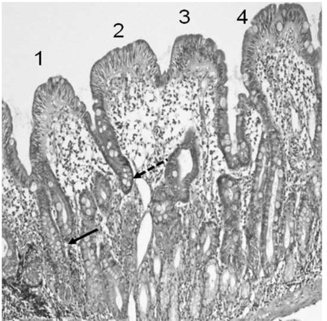
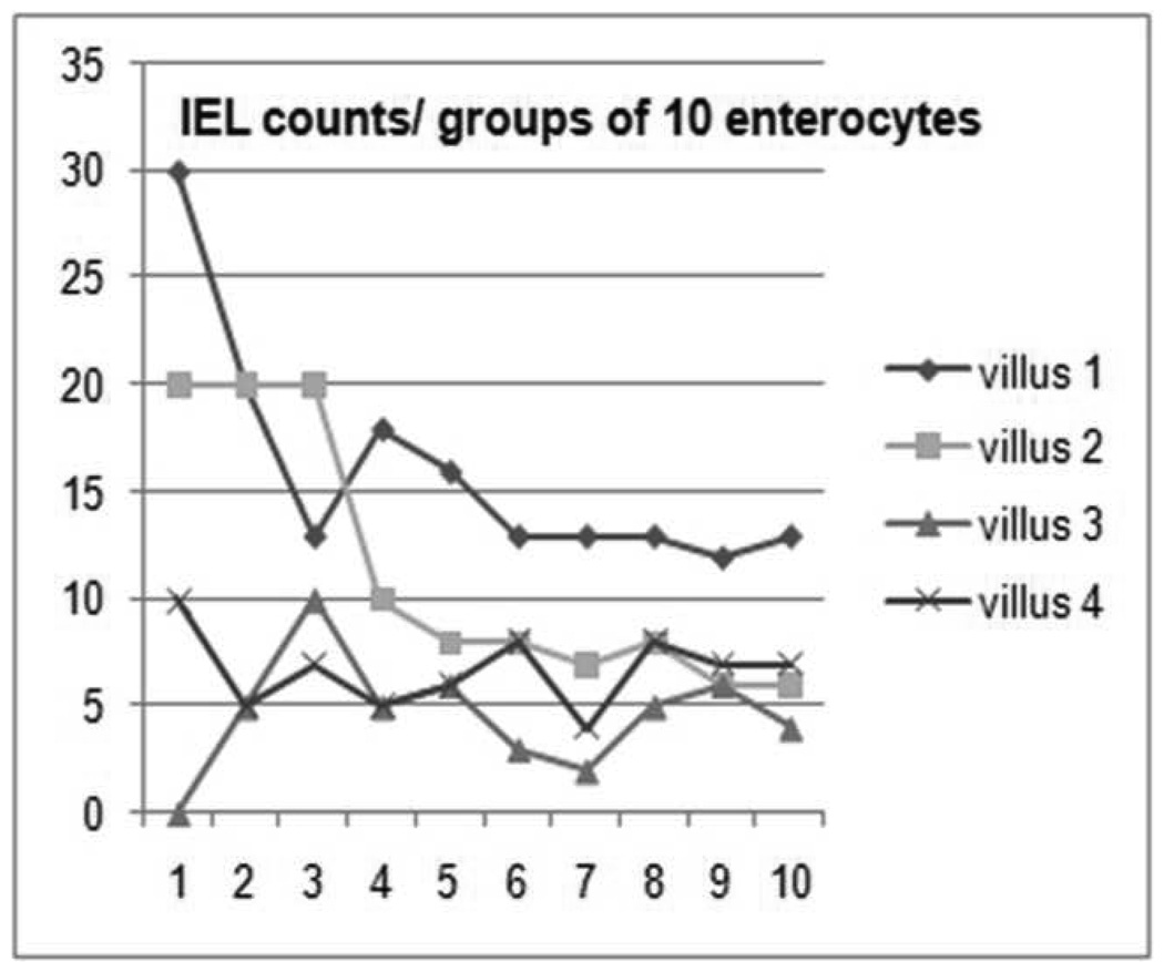
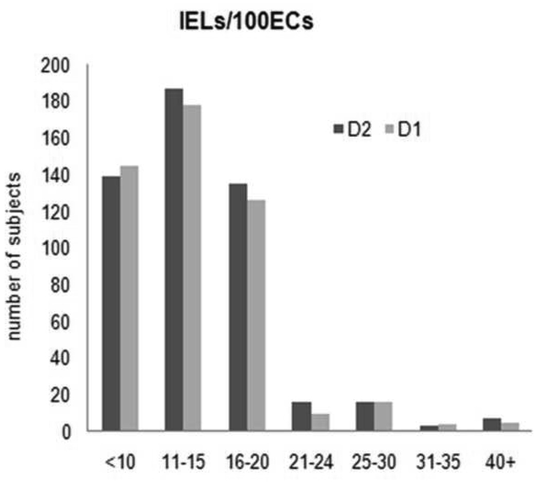
Methods: A random sample of an adult general population (n = 1000) was analyzed by upper endoscopy, duodenal biopsy, and serologic analysis of tissue transglutaminase (tTg) levels; endomysial antibody (EMA) levels were analyzed in samples that were tTg+. The cut off values for diagnosis of celiac disease were villous atrophy with 40 intraepithelial lymphocytes (IELs)/100 enterocytes (ECs).
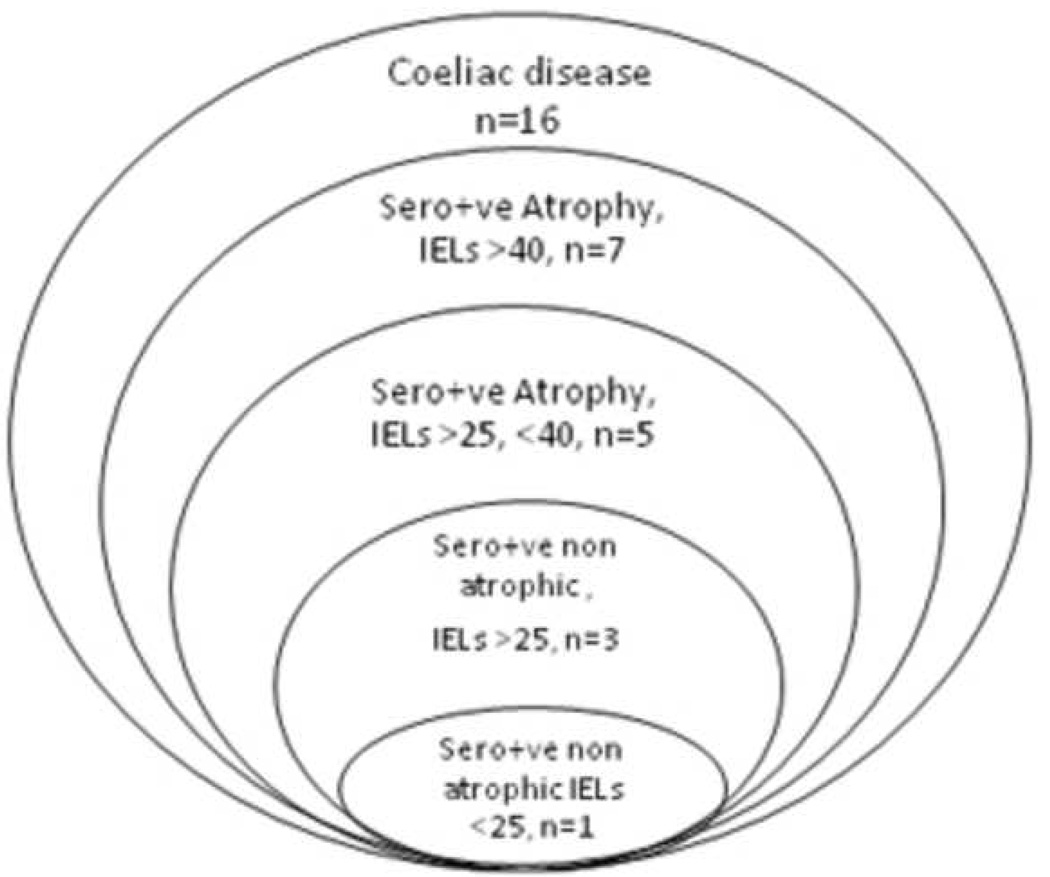
Results: Samples from 33 subjects were tTg+, and 16 were EMA+. Histologic analysis identified 7 of 1000 subjects (0.7%) with celiac disease; all were tTg+, and 6 of 7 were EMA+. Another 26 subjects were tTg+ (7/26 EMA+). This was addressed by a second quantitative pathology study (nested case control design) using a threshold of 25 IELS/100 ECs. In this analysis, all 13 samples that were tTg+ and EMA+ had > or =25 IELs/100 ECs. In total, 16 subjects (1.6%) had serologic and histologic evidence of gluten-sensitive enteropathy. IELs were quantified in duodenal biopsy samples from seronegative individuals (n = 500); 19 (3.8%) had >25 IELs and lymphocytic duodenosis.
Conclusions: Measurement of > or =25 IELs/100 ECs correlated with serologic indicators of celiac disease; a higher IEL threshold could miss 50% of cases. Quantification of tTg is a sensitive test for celiac disease; diagnosis can be confirmed by observation of > or =25 IELs/100ECs in duodenal biopsy specimens. Lymphocytic enteropathy (celiac disease and lymphocytic duodenosis) is common in the population (5.4%).
Copyright 2010 AGA Institute. Published by Elsevier Inc. All rights reserved.
Figures
References
-
- Dube C, Rostom A, Sy R, Cranney A, Saloojee N, Garritty C, Sampson M, Zhang L, Yazdi F, Mamaladze V, Pan I, Macneil J, Mack D, Patel D, Moher D. The prevalence of celiac disease in average-risk and at-risk Western European populations: a systematic review. Gastroenterology. 2005;128:S57–S67. - PubMed
-
- Murray JA, Van Dyke C, Plevak MF, Dierkhising RA, Zinsmeister AR, Melton LJ., 3rd Trends in the identification and clinical features of celiac disease in a North American community, 1950–2001. Clinical Gastroenterol Hepatol. 2003;1:19–27. - PubMed
-
- Talley NJ, Valdovinos M, Petterson TM, Carpenter HA, Melton LJ., 3rd Epidemiology of celiac sprue: a community-based study. Am J Gastroenterol. 1994;89:843–846. - PubMed
-
- Rossi TM, Albini CH, Kumar V. Incidence of celiac disease identified by the presence of serum endomysial antibodies in children with chronic diarrhea, short stature, or insulin-dependent diabetes mellitus. J Pediatr. 1993;123:262–264. - PubMed
-
- Stenhammar L, Ansved P, Jansson G, Jansson U. The incidence of childhood celiac disease in Sweden. J. Pediatr Gastroenterol Nutr. 1987;6:707–709. - PubMed
Publication types
MeSH terms
Substances
Grants and funding
LinkOut - more resources
Full Text Sources
Medical
Research Materials
