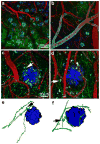
Multiphoton in vivo imaging of amyloid in animal models of Alzheimer's disease
- PMID: 20398680
- PMCID: PMC3117428
- DOI: 10.1016/j.neuropharm.2010.04.007
Multiphoton in vivo imaging of amyloid in animal models of Alzheimer's disease
Abstract
Amyloid-beta (Abeta) deposition is a defining feature of Alzheimer's disease (AD). The toxicity of Abeta aggregation is thought to contribute to clinical deficits including progressive memory loss and cognitive dysfunction. Therefore, Abeta peptide has become the focus of many therapeutic approaches for the treatment of AD due to its central role in the development of neuropathology of AD. In the past decade, taking the advantage of multiphoton microscopy and molecular probes for amyloid peptide labeling, the dynamic progression of Abeta aggregation in amyloid plaques and cerebral amyloid angiopathy has been monitored in real time in transgenic mouse models of AD. Moreover, amyloid plaque-associated alterations in the brain including dendritic and synaptic abnormalities, changes of neuronal and astrocytic calcium homeostasis, microglial activation and recruitment in the plaque location have been extensively studied. These studies provide remarkable insight to understand the pathogenesis and pathogenicity of amyloid plaques in the context of AD. The ability to longitudinally image plaques and related structures facilitates the evaluation of therapeutic approaches targeting toward the clearance of plaques.
Published by Elsevier Ltd.
Figures


Similar articles
-
Fibrillar Aβ triggers microglial proteome alterations and dysfunction in Alzheimer mouse models.Elife. 2020 Jun 8;9:e54083. doi: 10.7554/eLife.54083. Elife. 2020. PMID: 32510331 Free PMC article.
-
In Vivo Near-Infrared Two-Photon Imaging of Amyloid Plaques in Deep Brain of Alzheimer's Disease Mouse Model.ACS Chem Neurosci. 2018 Dec 19;9(12):3128-3136. doi: 10.1021/acschemneuro.8b00306. Epub 2018 Aug 16. ACS Chem Neurosci. 2018. PMID: 30067906
-
New Insights into the Spontaneous Human Alzheimer's Disease-Like Model Octodon degus: Unraveling Amyloid-β Peptide Aggregation and Age-Related Amyloid Pathology.J Alzheimers Dis. 2018;66(3):1145-1163. doi: 10.3233/JAD-180729. J Alzheimers Dis. 2018. PMID: 30412496
-
[Involvement of beta-amyloid in the etiology of Alzheimer's disease].Brain Nerve. 2010 Jul;62(7):691-9. Brain Nerve. 2010. PMID: 20675873 Review. Japanese.
-
Drebrin in Alzheimer's Disease.Adv Exp Med Biol. 2017;1006:203-223. doi: 10.1007/978-4-431-56550-5_12. Adv Exp Med Biol. 2017. PMID: 28865022 Review.
Cited by
-
Long-term in vivo imaging of β-amyloid plaque appearance and growth in a mouse model of cerebral β-amyloidosis.J Neurosci. 2011 Jan 12;31(2):624-9. doi: 10.1523/JNEUROSCI.5147-10.2011. J Neurosci. 2011. PMID: 21228171 Free PMC article.
-
APP transgenic mice: their use and limitations.Neuromolecular Med. 2011 Jun;13(2):117-37. doi: 10.1007/s12017-010-8141-7. Epub 2010 Dec 9. Neuromolecular Med. 2011. PMID: 21152995 Review.
-
In vivo imaging biomarkers in mouse models of Alzheimer's disease: are we lost in translation or breaking through?Int J Alzheimers Dis. 2010 Sep 30;2010:604853. doi: 10.4061/2010/604853. Int J Alzheimers Dis. 2010. PMID: 20953404 Free PMC article.
-
Potential Utility of Retinal Imaging for Alzheimer's Disease: A Review.Front Aging Neurosci. 2018 Jun 22;10:188. doi: 10.3389/fnagi.2018.00188. eCollection 2018. Front Aging Neurosci. 2018. PMID: 29988470 Free PMC article. Review.
-
Morphological and pathological evolution of the brain microcirculation in aging and Alzheimer's disease.PLoS One. 2012;7(5):e36893. doi: 10.1371/journal.pone.0036893. Epub 2012 May 16. PLoS One. 2012. PMID: 22615835 Free PMC article.
References
-
- Abramov AY, Canevari L, Duchen MR. Calcium signals induced by amyloid beta peptide and their consequences in neurons and astrocytes in culture. Biochim Biophys Acta. 2004b;1742:81–87. - PubMed
-
- Bacskai BJ, Hyman BT. Alzheimer’s disease: what multiphoton microscopy teaches us. Neuroscientist. 2002;8:386–390. - PubMed
-
- Bacskai BJ, Kajdasz ST, Christie RH, Carter C, Games D, Seubert P, Schenk D, Hyman BT. Imaging of amyloid-beta deposits in brains of living mice permits direct observation of clearance of plaques with immunotherapy. Nat Med. 2001;7:369–372. - PubMed
Publication types
MeSH terms
Substances
Grants and funding
LinkOut - more resources
Full Text Sources
Medical
Miscellaneous

