

Paramyxovirus assembly and budding: building particles that transmit infections
- PMID: 20398786
- PMCID: PMC2910131
- DOI: 10.1016/j.biocel.2010.04.005
Paramyxovirus assembly and budding: building particles that transmit infections
Abstract
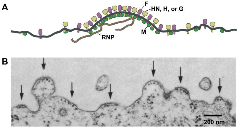
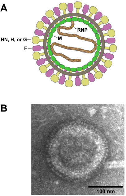
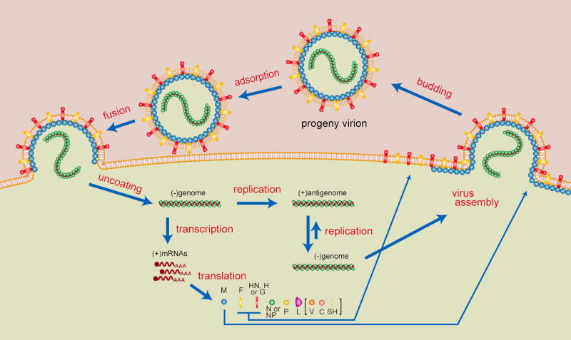
The paramyxoviruses define a diverse group of enveloped RNA viruses that includes a number of important human and animal pathogens. Examples include human respiratory syncytial virus and the human parainfluenza viruses, which cause respiratory illnesses in young children and the elderly; measles and mumps viruses, which have caused recent resurgences of disease in developed countries; the zoonotic Hendra and Nipah viruses, which have caused several outbreaks of fatal disease in Australia and Asia; and Newcastle disease virus, which infects chickens and other avian species. Like other enveloped viruses, paramyxoviruses form particles that assemble and bud from cellular membranes, allowing the transmission of infections to new cells and hosts. Here, we review recent advances that have improved our understanding of events involved in paramyxovirus particle formation. Contributions of viral matrix proteins, glycoproteins, nucleocapsid proteins, and accessory proteins to particle formation are discussed, as well as the importance of host factor recruitment for efficient virus budding. Trafficking of viral structural components within infected cells is described, together with mechanisms that allow for the selection of specific sites on cellular membranes for the coalescence of viral proteins in preparation of bud formation and virion release.
Copyright (c) 2010 Elsevier Ltd. All rights reserved.
Figures
References
-
- Alexander DJ. OIE Manual of Diagnostic Tests and Vaccines for Terrestrial Animals. Office of International Des Epizooties; Paris: 2009. Newcastle disease.
-
- Ali A, Nayak DP. Assembly of Sendai virus: M protein interacts with F and HN proteins and with the cytoplasmic tail and transmembrane domain of F protein. Virology. 2000;276:289–303. - PubMed
-
- Bieniasz PD. Late budding domains and host proteins in enveloped virus release. Virology. 2006;344:55–63. - PubMed
-
- Bishop KA, Broder CC. Hendra and Nipah: Lethal Zoonotic Paramyxoviruses. In: Scheld WM, Hammer SM, Hughes JM, editors. Emerging Infections. Washington, D. C: American Society for Microbiology; 2008. pp. 155–87.
Publication types
MeSH terms
Substances
Grants and funding
LinkOut - more resources
Full Text Sources
Other Literature Sources
