Review
. 2010 Aug;35(8):450-8.
doi: 10.1016/j.tibs.2010.03.002. Epub 2010 May 1.

The extended PP1 toolkit: designed to create specificity

Affiliations
Review

The extended PP1 toolkit: designed to create specificity

Mathieu Bollen et al. Trends Biochem Sci. 2010 Aug.

Abstract

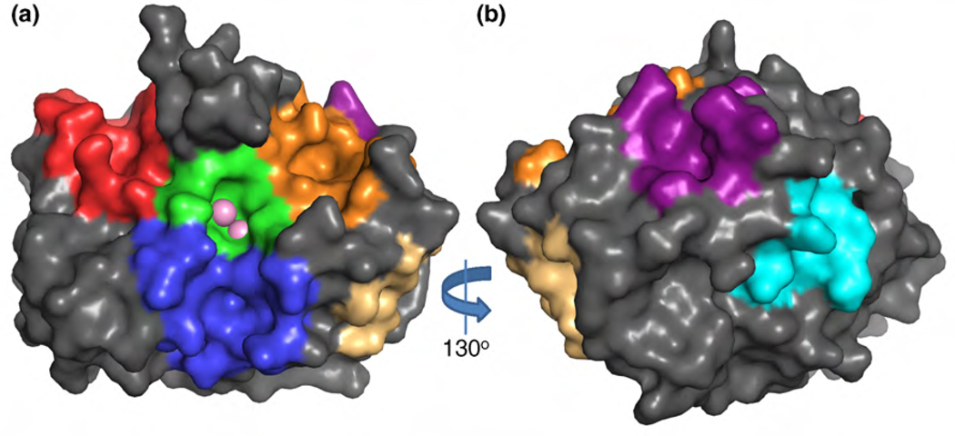
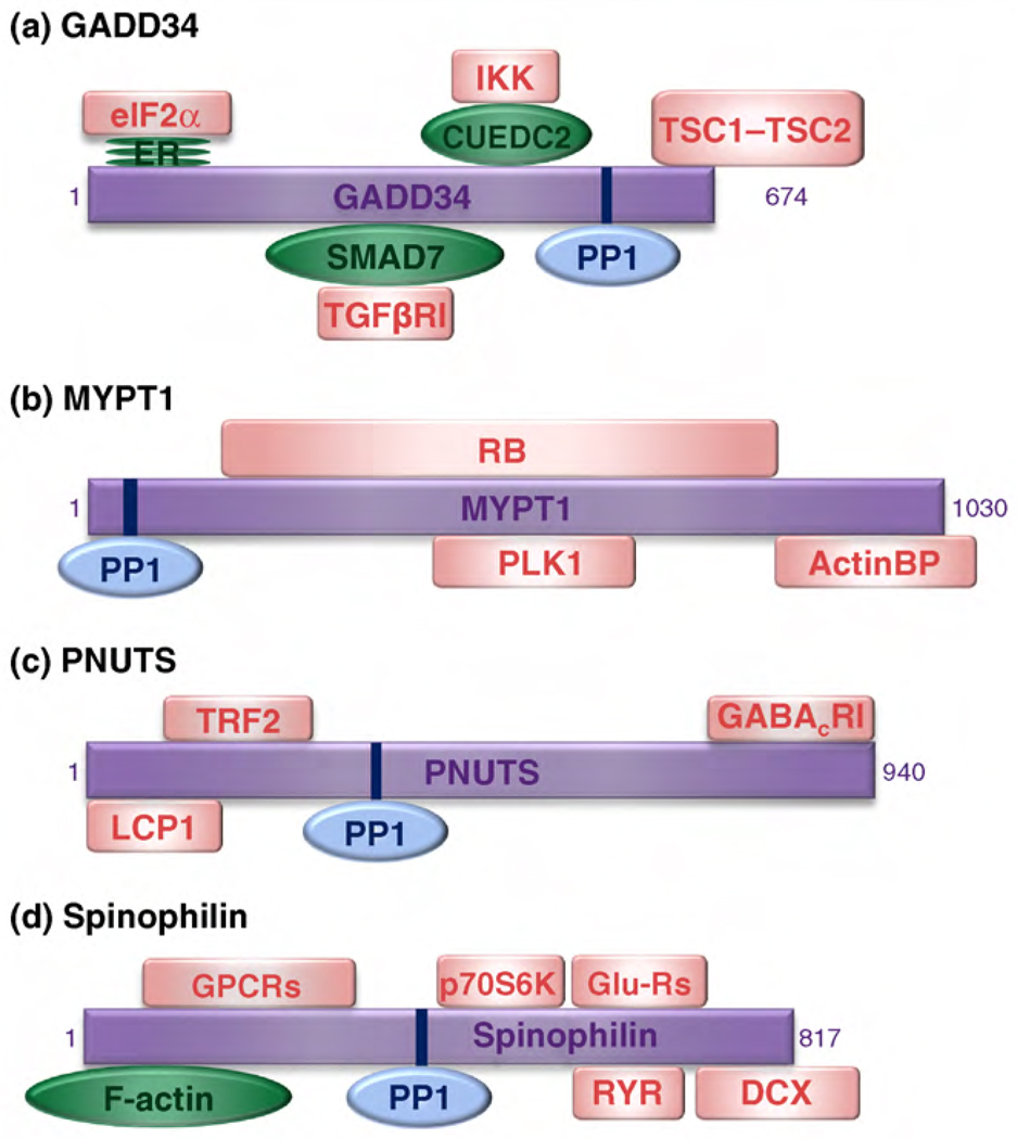
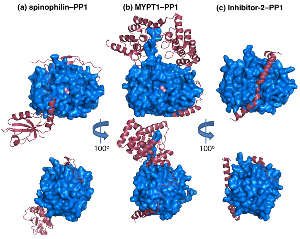
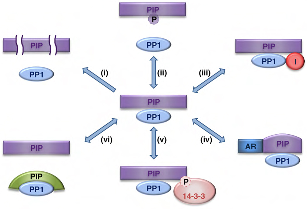
Protein Ser/Thr phosphatase-1 (PP1) catalyzes the majority of eukaryotic protein dephosphorylation reactions in a highly regulated and selective manner. Recent studies have identified an unusually diversified PP1 interactome with the properties of a regulatory toolkit. PP1-interacting proteins (PIPs) function as targeting subunits, substrates and/or inhibitors. As targeting subunits, PIPs contribute to substrate selection by bringing PP1 into the vicinity of specific substrates and by modulating substrate specificity via additional substrate docking sites or blocking substrate-binding channels. Many of the nearly 200 established mammalian PIPs are predicted to be intrinsically disordered, a property that facilitates their binding to a large surface area of PP1 via multiple docking motifs. These novel insights offer perspectives for the therapeutic targeting of PP1 by interfering with the binding of PIPs or substrates.


Figures

Figure 1
Figure 1
Surface representation of the structure of PP1α (PDB ID 1FJM). (a) The active site of PP1 (green) contains two metal ions (pink spheres) and lies at the Y-shaped intersection of three substrate-binding grooves; the hydrophobic (blue), the acidic (orange), and the C-terminal (red). (b) A 130° rotation of a, showing binding sites for the PP1-docking motifs RVxF (purple), SILK (cyan) and MyPhoNE (wheat).
Figure 2
Figure 2
PP1-interacting proteins with multiple substrate-targeting domains. The figure shows selected PIPs that can act as signal integrators because they have multiple substrate-targeting domains. (a) GADD34, (b) MYPT1, (c) PNUTS and (d) spinophilin target PP1 (blue) either directly to its substrates (pink squares) or via adaptor proteins (green ovals). Some of the substrates (LCP1, TRF2, GABACR1 and GPCRs) are still hypothetical. Abbreviations: ER, endoplasmic reticulum; RB, retinoblastoma protein; ActinBP, actin-binding proteins (including myosin, merlin and moesin); GPCRs, G protein-coupled receptors; Glu-Rs, glutamate receptors; RYR, ryanodine receptors.
Figure 3
Figure 3
Crystal structures of protein–PP1 complexes. The regulatory proteins are shown as pink ribbons and PP1 as a blue surface representation. Top images are centered around the active site of PP1 whereas the bottom images have been rotated 100° to highlight the interaction within the RVxF docking motif. (a) The crystal structure of spinophilin417–583–PP1α7–330 (PDB ID 3EGG). (b) The crystal structure of MYPT11–299–PP1δ (PDB ID 1S70). (c) The crystal structure of Inhibitor-2–PP1γ (PDB ID 2O8A). These PP1 interacting proteins share only the common RVxF motif interaction. All other interactions are unique for each holoenzyme.
Figure 4
Figure 4
PIP-mediated regulation of PP1 holoenzymes. The figure gives an overview of regulatory mechanisms that affect PIPs, and hence PP1 function. (i) Controlled proteolysis of PIPs. (ii) Dissociation of holoenzymes by the phosphorylation of PIPs. (iii) Recruitment or dissociation of inhibitory PIPs. (iv) Allosteric regulation by the binding of metabolites or proteins to PIPs. (v) Substrate masking by 14-3-3 protein binding. (vi) Competition between PIPs for the same binding sites on PP1. Abbreviations: AR, allosteric regulator; I, inhibitor; P, phosphate.

Similar articles

Cited by

References

    1. Olsen JV, et al. Quantitative phosphoproteomics reveals widespread full phosphorylation site occupancy during mitosis. Sci. Signal. 2010;3:ra3. - PubMed
    1. Ubersax JA, Ferrell JE., Jr Mechanisms of specificity in protein phosphorylation. Nat. Rev. Mol. Cell Biol. 2007;8:530–541. - PubMed
    1. Manning G, et al. Evolution of protein kinase signaling from yeast to man. Trends Biochem. Sci. 2002;27:514–520. - PubMed
    1. Moorhead GB, et al. Emerging roles of nuclear protein phosphatases. Nat. Rev. Mol. Cell Biol. 2007;8:234–244. - PubMed
    1. Virshup DM, Shenolikar S. From promiscuity to precision: protein phosphatases get a makeover. Mol. Cell. 2009;33:537–545. - PubMed

Publication types

Substances