. 2010 Mar 14;18(4):436-48.
doi: 10.1016/j.str.2010.01.013.

Structure of concatenated HAMP domains provides a mechanism for signal transduction

Affiliations

Structure of concatenated HAMP domains provides a mechanism for signal transduction

Michael V Airola et al. Structure. .

Abstract

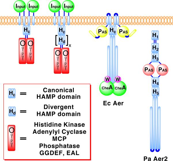
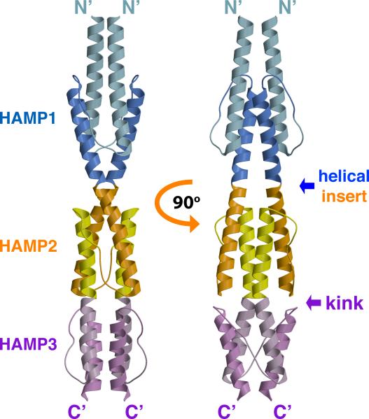
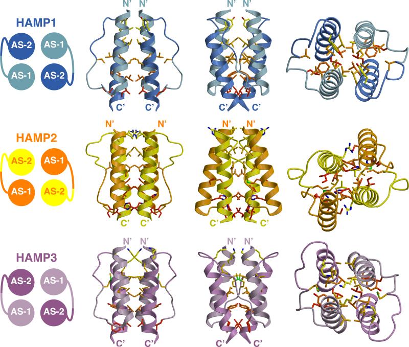
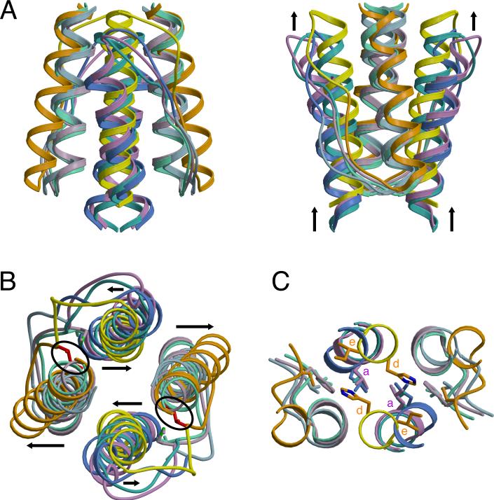
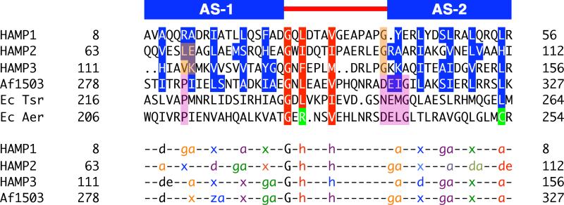
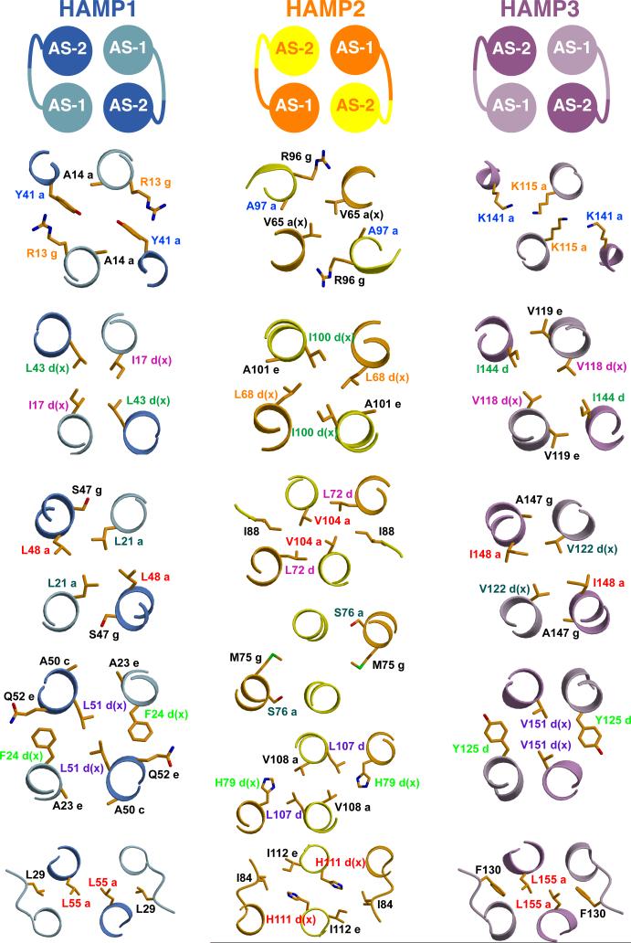
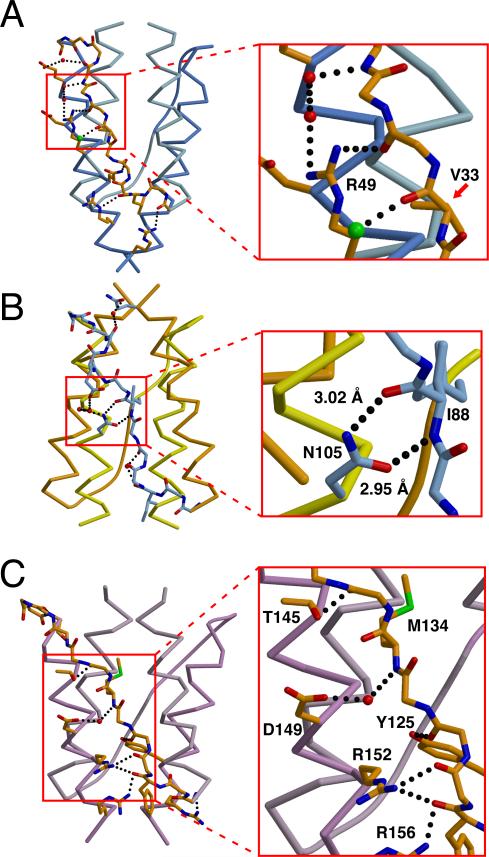
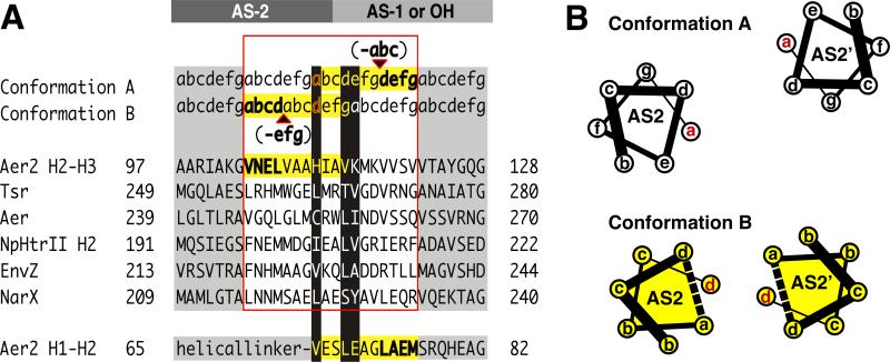
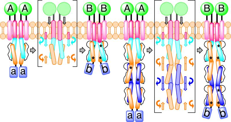
HAMP domains are widespread prokaryotic signaling modules found as single domains or poly-HAMP chains in both transmembrane and soluble proteins. The crystal structure of a three-unit poly-HAMP chain from the Pseudomonas aeruginosa soluble receptor Aer2 defines a universal parallel four-helix bundle architecture for diverse HAMP domains. Two contiguous domains integrate to form a concatenated di-HAMP structure. The three HAMP domains display two distinct conformations that differ by changes in helical register, crossing angle, and rotation. These conformations are stabilized by different subsets of conserved residues. Known signals delivered to HAMP would be expected to switch the relative stability of the two conformations and the position of a coiled-coil phase stutter at the junction with downstream helices. We propose that the two conformations represent opposing HAMP signaling states and suggest a signaling mechanism whereby HAMP domains interconvert between the two states, which alternate down a poly-HAMP chain.


Figures

Figure 1
Figure 1. Domain Architectures of Representative HAMP Containing Proteins
Schematic depicting the location of canonical HAMP domains and poly-HAMP chains in transmembrane and soluble signaling proteins. Poly-HAMP chains, [Hd]x, can extend from 2 to 31 consecutive HAMP domains. EcAer is shown binding to the histidine kinase CheA and coupling protein CheW, proteins PaAer2 also likely interacts with.
Figure 2
Figure 2. Crystal Structure of the Aer2 N-Terminal Domain Contains Three Successive and Interwoven HAMP Domains
Ribbon presentation of the Aer2 (1-172) dimer with HAMP1 (AS1: light blue, AS2: blue), HAMP2 (AS1: orange, AS2: yellow), and HAMP3 (AS1: light purple, AS2: purple). HAMP2/3 forms a concatenated structure. AS2 of HAMP1 is contiguous with AS1 of HAMP2 and AS2 of HAMP2 is contiguous with AS1 of HAMP3. HAMP3 is rotated roughly 90 relative to HAMP1 and HAMP2.
Figure 3
Figure 3. Structures of the Aer2 HAMP Domains
Side and top views (90° rotation) of individual Aer2 HAMP domains with buried side chains (Carbon: yellow to red, Nitrogen: blue, Sulfur: green). HAMP1 and HAMP3 sidechains are in-register and HAMP2 sidechains are offset. HAMP2 has an unusual trapezoidal shape (as viewed from the side) and rhombic arrangement of helices in cross section. The position of the connector and hydrophobic residue 2 (HR2) correlates with changes in helical register, helix crossing angles, and helical rotation. I88 (HR2, HAMP2) inserts between AS1 and AS2, and V33 (HR2, HAMP1) and M134 (HR2, HAMP3) pack against the periphery of AS1 and AS2.
Figure 4
Figure 4. Superposition of HAMP Structures Reveals Two Distinct Conformations for Providing Different Signals to Downstream Domains
Ribbon presentation of HAMP superpositions in (A) side and (B) top views displaying differences in helix orientation between overlapping HAMP1, HAMP3, and Af1503 (AS1: light green, AS2: turquoise) helices and distinct conformation of HAMP2 (AS1: orange, AS2: yellow) with offset helices, change in helix crossing angles (black arrows), and the inserted HR2 (I88: red) residue (black circle). Note the vertical displacement of AS2 relative to AS1. (C) Distal region of HAMP domains where signal output is directly linked to the conformation of AS2. His111 (HAMP2) occupies a typical “d” position, with the analogous residue in other HAMPs (L55: HAMP1, L155: HAMP3, L326: Af1503) holding an “a” position, in which they interact with hydrophobic residue 1 (HR1) of the connector.
Figure 5
Figure 5. Residue Conservation and Packing Modes of HAMP Domains
Sequence alignment of representative HAMP domains: Aer2, Af1503, EcTsr, and EcAer, showing conservation of the buried core (blue) and connector motif (red) residues (top panel). The bottom panel denotes the heptad position of core residues (a, d, e, g, z = undefined, x = directed at supercoil axis), as defined by TWISTER, in a variety of packing modes: a-d, x-da (x-ga or x-de), and x-x. Sets of interacting residues are color-coded to highlight the sidechains of HAMP1, HAMP3, and Af1503 (in-register) and HAMP2 (offset) (bottom panel). “G” and “h” correspond to conserved residues of the connector motif and are also color coded with the layer of interacting residues. Position of the conserved proline residue (purple) in canonical HAMP domains corresponds to residues that occupy different heptad positions (peach) in alternative HAMP conformations. Aer2 HAMPs conserve Gly residues (peach) at the beginning of AS2, which allows close association of domains in a poly-HAMP chain, compared to the DExG motif of canonical HAMPs (purple).
Figure 6
Figure 6. Sidechain Interactions of Aer2 HAMP domains
Cross-sections of the Aer2 HAMP domains displaying a variety of sidechain packing modes: a-d, x-da (x-ga or x-de), and x-x. Clockwise and counterclockwise rotation of AS1 and AS2 alters the corresponding sidechain positions (color-coded) in HAMP2 compared to HAMP1 and HAMP3. These changes generate a greater helix-helix separation that increases the overall solvent accessibility of HAMP2 residues at the inter-helix interface (Figure S3). Sidechains of HAMP2 do not occupy the same plane due to a change in helical register; they are shown with the nearest layer. Helices are viewed with the N-terminus pointing toward the viewer.
Figure 7
Figure 7. Hydrogen Bonding Interactions Between the Connector and Helices Stabilize the Aer2 HAMP Domains
Variable hydrogen bonding interactions and associations between the connector (peptide backbone) and helices (Cα trace) stabilize the different conformations of (A) HAMP1, (B) HAMP2, and (C) HAMP3. N105 hydrogen bonds (dotted line) with the peptide backbone to stabilize the position of the inserted I88 residue (hydrophobic residue 2: HR2) in HAMP2 (inset B). R49 forms a hydrogen bonding network with the connector peptide backbone and two water molecules (red). Peptide backbone of V33 (HR2) in HAMP1 forms a salt bridge with a chlorine atom (green) (inset A). D149 of HAMP3 forms a salt bridge with a water molecule (red) to the amide nitrogen of M134 (HR2) (inset C).
Figure 8
Figure 8. Output Mechanism Involving “Stutter Compensation”
(A) Model for signal transfer that shifts the position of a stutter on either side of the AS2 junction with AS1 (poly-HAMPs) or output helices (OH) (canonical HAMPs). Helical junctions are shown for representative HAMP domains and Aer2 HAMP 2-3. Stutters (bold) are defined as either an insertion of four residues or a deletion of three residues in the heptad repeat (a-g) and effect adjacent residues (yellow highlight). Residues at the junction switch heptad positions (black highlight) and are compatible with different heptad positions. This model predicts that Aer2 HAMP2 contains a stutter (shown in bold) in AS1 (bottom sequence). Whereas the heptad assignments in HAMP2 AS-1 are as shown, the splayed helices of AS-1 do not generate a true hendecad repeat but do contain an elongated i to i+4 (E66-A70) hydrogen bond distance (3.5 Å) typical of stutters. (B) Helical wheel diagrams of AS2 helices in conformation A (HAMP1/3) and B (HAMP2) highlighting the helical rotation and translation associated with a stutter (dotted line) in AS2.
Figure 9
Figure 9. Model for HAMP Domain Signal Transduction
Schematic of a ligand dependent, two-state (A and B) HAMP domain signal transduction model highlighting the movement and rotation of helices in a canonical HAMP system and a poly-HAMP system. In the poly-HAMP system successive AS1 helices are contiguous with the preceding AS2 helices. HAMP domain structural rearrangements correspond to a change in helical register, helix-helix crossing angle, and helix rotation. Asterisks denote stutters within HAMP domains. Signal input is shown as a symmetric and downward piston-like displacement of transmembrane helix 2 (TM2), but both an asymmetric displacement or a helical rotation of TM2 would likely lead to the same conformational changes within HAMP domains.

Similar articles

Cited by

References

    1. Altschul SF, Madden TL, Schaffer AA, Zhang JH, Zhang Z, Miller W, Lipman DJ. Gapped BLAST and PSI-BLAST: a new generation of protein database search programs. Nucleic Acids Research. 1997;25:3389–3402. - PMC - PubMed
    1. Ambroggio XI, Kuhlman B. Design of protein conformational switches. Current Opinion in Structural Biology. 2006;16:525–530. - PubMed
    1. Ames P, Zhou Q, Parkinson JS. Mutational analysis of the connector segment in the HAMP domain of Tsr, the Escherichia coli serine chemoreceptor. Journal of Bacteriology. 2008;190:6676–6685. - PMC - PubMed
    1. Appleman JA, Chen LL, Stewart V. Probing conservation of HAMP linker structure and signal transduction mechanism through analysis of hybrid sensor kinases. Journal of Bacteriology. 2003;185:4872–4882. - PMC - PubMed
    1. Aravind L, Ponting CP. The cytoplasmic helical linker domain of receptor histidine kinase and methyl-accepting proteins is common to many prokaryotic signalling proteins. Fems Microbiology Letters. 1999;176:111–116. - PubMed

Publication types

LinkOut - more resources