Review
. 2010 Apr 15;66(1):15-36.
doi: 10.1016/j.neuron.2010.01.018.

Design principles of insect and vertebrate visual systems

Affiliations
Review

Design principles of insect and vertebrate visual systems

Joshua R Sanes et al. Neuron. .

Abstract

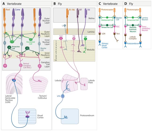
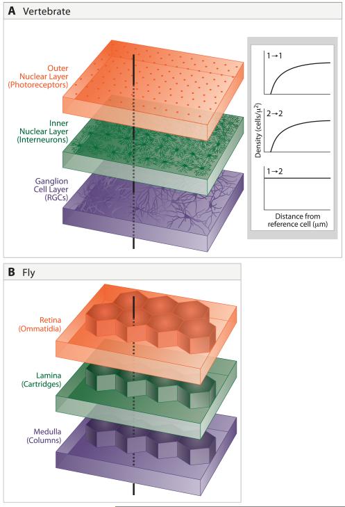
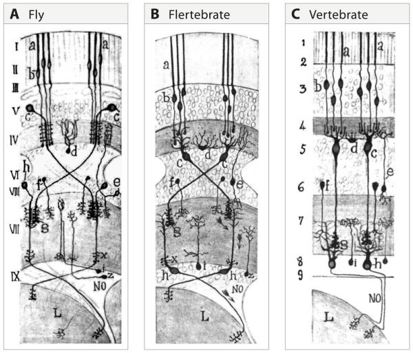
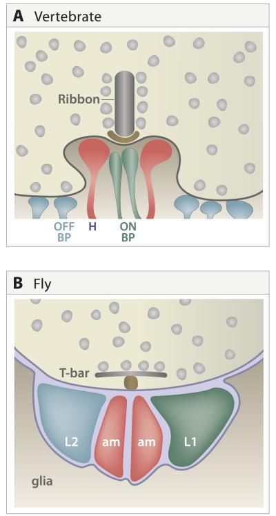
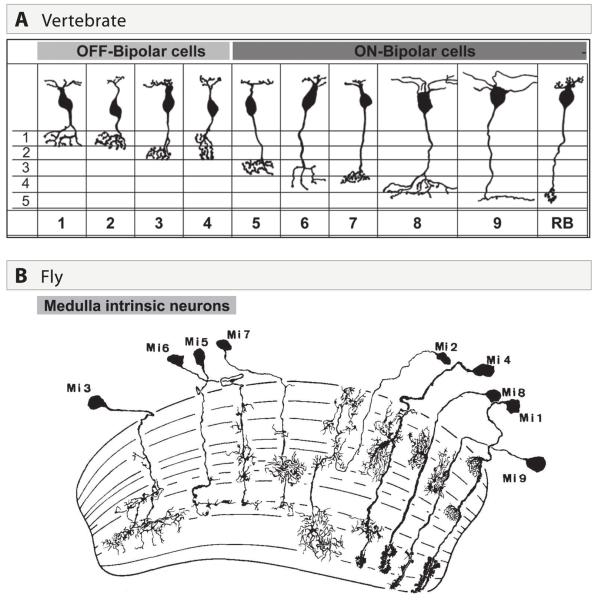
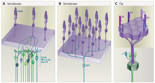
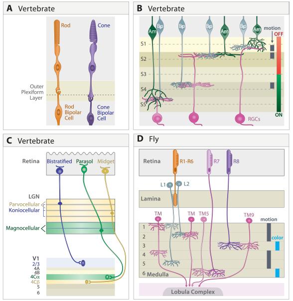
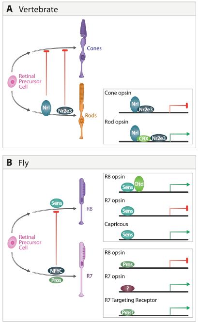
A century ago, Cajal noted striking similarities between the neural circuits that underlie vision in vertebrates and flies. Over the past few decades, structural and functional studies have provided strong support for Cajal's view. In parallel, genetic studies have revealed some common molecular mechanisms controlling development of vertebrate and fly visual systems and suggested that they share a common evolutionary origin. Here, we review these shared features, focusing on the first several layers-retina, optic tectum (superior colliculus), and lateral geniculate nucleus in vertebrates; and retina, lamina, and medulla in fly. We argue that vertebrate and fly visual circuits utilize common design principles and that taking advantage of this phylogenetic conservation will speed progress in elucidating both functional strategies and developmental mechanisms, as has already occurred in other areas of neurobiology ranging from electrical signaling and synaptic plasticity to neurogenesis and axon guidance.


Figures

Figure 1
Figure 1. Structures underlying the first stages of visual processing
A. Mammalian visual system, showing retina, dorsal lateral geniculate nucleus (LGN), superior colliculus (called optic tectum in lower vertebrates) and primary visual cortex (also called Area 17, striate cortex, or V1). Main retinal cell types are indicated. B. Drosophila visual system, showing retina, lamina, medulla, and the lobula complex, which comprises the lobula and lobula plate. A few cell types are shown. C,D. Similar steps in transfer of information through early stages of vertebrate and Drosophila visual systems.
Figure 2
Figure 2. Laminar and radial arrangement of cells and connections
A. Cells are arranged in mosaics in the outer and inner nuclear and ganglion cell layers of the vertebrate retina. Because of mosaic spacing, a line drawn perpendicular to the layers will intersect the dendritic field of at least one representative of each cell type. Inset shows distribution of cells within two amacrine mosaics, redrawn from Rockhill et al. (2000). For each cell type, spacing is non-random, with fewer near neighbors than would be expected by chance. However, mosaics are independent of each other, so distances between cells of types 1 and 2 are randomly distributed. B. In Drosophila, spatial relationships among successive stages are strictly determined rather than probabilistic. Retinal ommatidia overly laminar cartridges, which in turn overly medullary columns.
Figure 3
Figure 3. Cajal recognized the similarity of fly and vertebrate visual systems
A. The retina (I – III), lamina (IV and V) and medulla (VI – VIII) and lobula region (L) of the fly visual system. The somas appear in their natural position. a, b. photoreceptor; c, lamina monopolar neuron; h, transmedullary neuron. B. In this drawing of the insect visual system, Cajal “moved” the cell bodies to correspond to their positions in vertebrates, without changing the positions of their synaptic contacts. We refer to this as the “Flertebrate” arrangement. c, lamina monopolar neurons take on the appearance of bipolar neurons (see “c” in right panel); d, amacrine cells in the fly appear as horizontal cells (see “d” in right panel). h, transmedullary cells appear as retinal ganglion cells (see “h” in right panel). C. Schematic of the main cell types in the vertebrate retina and their connections. The Arabic numerals indicate regions of the vertebrate retina that Cajal viewed as similar to the corresponding layers marked with Roman numerals in the left panel. (From Cajal and Sanchez, 1915; adapted from Meinertzhagen, 1993).
Figure 4
Figure 4. Multiple contact synapses
A. Portion on a cone pedicle in the outer plexiform layer, showing its synapses with processes of multiple horizontal, ON bipolar and OFF bipolar cells. Ribbon, structure at the active zone of these synapses specialized for tonic transmitter release. B. Tetrad in Drosophila lamina, showing synapses of photoreceptor axon on processes of L1, L2 and amacrine (am) cell dendrites. Like the vertebrate photoreceptor, that of Drosophila contains an unusually large presynaptic specialization, in this case a T-bar (T). Schematic is simplified to show all postsynaptic elements in the same plane; L1 and L2 are paired equatorially while the am elements are paired in polar positions. In some cases, one amacrine cell process is replaced with a process from L3.
Figure 5
Figure 5. Diversification of main neuronal types into multiple subtypes
A. Comprehensive inventory of bipolar cell subtypes in mouse retina (from Wassle et al. 2009). B. Comprehensive inventory of medulla intrinsic neuron subtypes in Drosophila (from Fischbach and Dittrich et al. 1989).
Figure 6
Figure 6. Divergent and convergent connections of photoreceptors
A. A single mouse cone connects with at least one of each of the 10 cone bipolar cell types. B. A single mouse cone bipolar (BP1 sketched here) receives input from at least 10 cones. C. A single lamina neuron cartridge receives input from a single cell of each R1-R6 class, each in a different ommatidium. All six R cells synapse on the L1-L3 neurons associated with the cartridge.
Figure 7
Figure 7. Parallel processing of visual features in distinct laminae
A. Rod and cone terminals occupy distinct sublaminae in the vertebrate outer plexiform layer, where they synapse on dendrites of rod and cone bipolars, respectively. In some species (for example, chicks) the cone sublamina is further subdivided. B. Axons of ON bipolar cells form terminals in the inner part of the vertebrate inner plexiform layer, where they form synapses on ON RGCs, whose dendrites arborize in the same zone. Likewise, OFF bipolar axons and OFF RGC dendrites arborize and form synapses in an outer zone. Bipolar, amacrine and retinal ganglion cells are further subdivided into groups that form lamina-specified connections underlying sensitivity to other visual features; sublaminae specialized for processing directional motion in mouse are indicated. C. In primates, distinct sets of RGCs form the magnocellular, parvocellular and koniocellular pathways. Although their functions are overlapping, they are sometimes viewed as being specialized for processing information about motion, form, and color, respectively. Synapses in these pathways, are segregated into distinct laminae in LGN and project to distinct targets in primary visual cortex. D. In Drosophila, R1-R6 are critical for sensitivity to motion while R7 and R8 are required for color. These two classes of photoreceptors project directly (R7,8) or indirectly (R1-6, via the lamina) to separate layers in the medulla. Layers specialized to process information about color and motion are indicated. Only the distal 6 layers of the medulla are shown.
Figure 8
Figure 8. Transcriptional control of photoreceptor diversification
A. Regulation of photoreceptor subtype identity in the mouse retina. Nrl and Nr2e3 promote rod development and repress cone cell programs in rods. These transcription factors also regulate photoreceptor subtype-specific expression of opsins in rods and are likely to regulate other genes controlling rod and cone cell development (adapted from Onishi et al. 2009). B. Regulation of photoreceptor subtype in fly retina. NFYC represses R8 development in R7 by preventing Senseless expression. Senseless regulates R8 and R7 subtype-specific opsin expression and has also been implicated in expression of Capricious and other genes regulating R8 targeting. Prospero also regulates opsin expression and acts in parallel with other transcription factors to regulate R7 targeting. R7 and R8 subtypes can be further divided by the opsins they express (see text).
Figure 9
Figure 9. Dscam proteins mediates homotypic repellent interactions required for tiling of visual interneurons
A. Dscam is expressed in several amacrine cell subsets in mouse retina. In mutants lacking DscamSCAM, processes of these cells fasciculate and, in consequence, their mosaic arrangement is perturbed. Other amacrine subtypes, which did not express Dscam, are unaffected. B. Dscam2 is required selectively in L1 neurons for tiling in the medulla. Terminals of L1 neurons lacking Dscam2 extend into neighboring cartridges. Tiling of L2 axon terminals is normal in Dscam2 mutants.
Figure 10
Figure 10. Formation of lamina-specific connections
A. Inner plexiform layer. Processes of many amacrine cells ramify from the outset in appropriate sublaminae. Development of RGC dendrites exhibits subtype-specific variations: arbors of some types are initially diffuse then remodel, other are lamina-restricted from an early stage, and still others develop in discrete steps. The first two are shown here. Bipolar cells are born and extend axons late; their processes initially span multiple sublaminae, then become lamina-restricted. B. Lateral geniculate nucleus. Axons from the ipsi- and contralateral eyes initially arborize broadly. Activity-dependent processes promote refinement to appropriate target laminae. C. Fly lamina. The growth cones of R1-6 axons from the same ommatidium initially terminate in a tight cluster in a temporary layer bounded by glia. Interactions between growth cones promote extension away from one another in defined orientations where each projects to a different set of lamina neurons in surrounding cartridges and forms synapses with them. Some of the molecules implicated in discrete steps are indicated. D. Fly medulla. The axons of R7, R8, L3 and L5 exhibit cell type-specific behaviors as they form lamina-specific terminals in the medulla.
Figure 11
Figure 11. Homophilic interactions promote lamina-specific arborization and synapse formation
A. In chick retina, non-overlapping subsets of interneurons and RGCs express one of four related immunoglobulin superfamily molecules: Sidekick 1, Sidekick 2, Dscam, and DscamL. Most of the pre- and postsynaptic cells expressing the same gene arborize in a distinct subset of inner plexiform sublaminae. Loss and gain of function studies support the idea that these genes promote lamina-specific synapse formation; as an example, sketches show results from manipulating Sidekick 1 levels in RGCs. Recognition molecules regulating the targeting of other subtypes (green) have not been identified. B. In fly, N-cadherin and Capricious affect lamina-specific targeting of R7 and R8 terminals to appropriate medullar laminae. It is not known whether lamina-specific targeting requires expression of N-cadherin on TM5 and Capricious on TM9.

References

    1. Agathocleous M, Harris WA. From progenitors to differentiated cells in the vertebrate retina. Annu Rev Cell Dev Biol. 2009;25:45–69. - PubMed
    1. Andreae LC, Lumsden A, Gilthorpe JD. Chick Lrrn2, a novel downstream effector of Hoxb1 and Shh, functions in the selective targeting of rhombomere 4 motor neurons. Neural Dev. 2009;4:27. - PMC - PubMed
    1. Badea TC, Cahill H, Ecker J, Hattar S, Nathans J. Distinct roles of transcription factors brn3a and brn3b in controlling the development, morphology, and function of retinal ganglion cells. Neuron. 2009;61:852–864. - PMC - PubMed
    1. Barlow HB, Hill RM, Levick WR. Retinal Ganglion Cells Responding Selectively to Direction and Speed of Image Motion in the Rabbit. J Physiol. 1964;173:377–407. - PMC - PubMed
    1. Bazigou E, Apitz H, Johansson J, Loren CE, Hirst EM, Chen PL, Palmer RH, Salecker I. Anterograde Jelly belly and Alk receptor tyrosine kinase signaling mediates retinal axon targeting in Drosophila. Cell. 2007;128:961–975. - PubMed

Publication types