Review
. 2010 Aug 15;344(2):543-54.
doi: 10.1016/j.ydbio.2010.04.010. Epub 2010 Apr 23.

Cranial neural crest migration: new rules for an old road

Affiliations
Review

Cranial neural crest migration: new rules for an old road

Paul M Kulesa et al. Dev Biol. .

Abstract

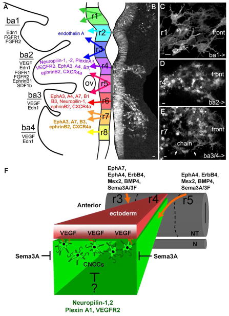
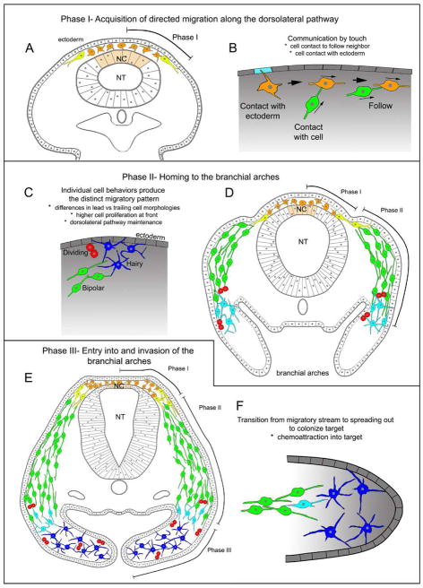
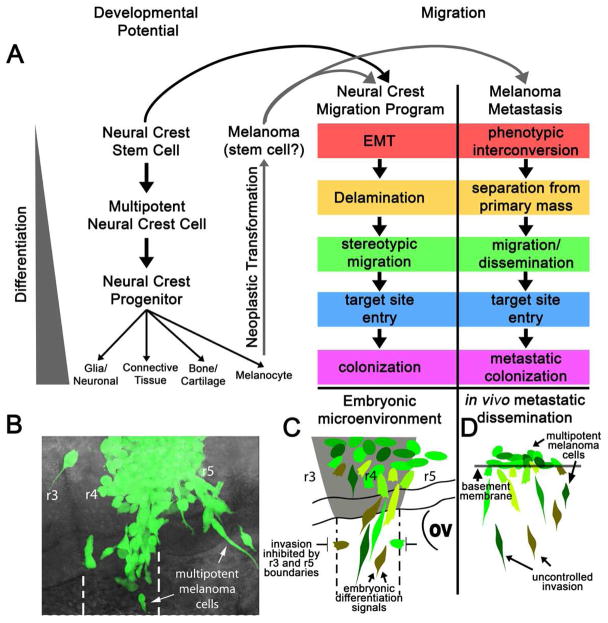
The neural crest serve as an excellent model to better understand mechanisms of embryonic cell migration. Cell tracing studies have shown that cranial neural crest cells (CNCCs) emerge from the dorsal neural tube in a rostrocaudal manner and are spatially distributed along stereotypical, long distance migratory routes to precise targets in the head and branchial arches. Although the CNCC migratory pattern is a beautifully choreographed and programmed invasion, the underlying orchestration of molecular events is not well known. For example, it is still unclear how single CNCCs react to signals that direct their choice of direction and how groups of CNCCs coordinate their interactions to arrive at a target in an ordered manner. In this review, we discuss recent cellular and molecular discoveries of the CNCC migratory pattern. We focus on events from the time when CNCCs encounter the tissue adjacent to the neural tube and their travel through different microenvironments and into the branchial arches. We describe the patterning of discrete cell migratory streams that emerge from the hindbrain, rhombomere (r) segments r1-r7, and the signals that coordinate directed migration. We propose a model that attempts to unify many complex events that establish the CNCC migratory pattern, and based on this model we integrate information between cranial and trunk neural crest development.


Figures

Figure 1
Figure 1. The cranial neural crest cell migratory pattern; cellular features and signaling pathways
(A) A schematic representation showing recently discovered key guidance cues involved in CNCC migration. (B) The cranial NCCs migrate in 3 distinct streams as seen by membrane (Gap43-GFP) and nuclear (H2B-mCherry) labeling (introduced into premigratory NCCs by electroporation delivery) reduced to grayscale for clarity. (C) CNCCs that emerge from mid-r3 and more rostral migrate in a broad wave and display multiple filopodial protrusions. (D) CNCCs that emerge mid-r3 to mid-r5 are sculpted into a tight stream adjacent to r4 that spreads out at the front (E) Post-otic NCCs that emerge from mid-r5 and more caudal migrate as an initial wave, followed by NCCs that form chain-like arrays. The arrows point to cells that travel in a chain-like array. (F) A schematic representation of the molecules guiding the r4 NCC stream. The r4 NCCs express neuropilins, Plexin A1 and VEGFR2. The overlaying ectoderm expresses VEGF, which is a NCC chemoattractant. R3 and r5 secrete semaphorin3A, which is a NCC inhibitor. A guidance cue that prevents the r4 NCCs migrating ventromedially is as yet unknown (?). r, rhombomere; ba, branchial arch; OV, otic vesicle, NT, neural tube, N, notochord. The scale bars are 20um in (B) and 10um in (C–E).
Figure 2
Figure 2. The three phases of cranial neural crest cell migration and characteristic cell behaviors. Phase 1: Acquisition of directed migration along the dorsolateral pathway
(A) Cranial NCC migration starts around Hamburger and Hamilton (HH) Stage 11 in chick. During Phase I, the lead cranial NCCs emerge from the dorsal neural tube (beige). Initially the cranial NCCs do not exhibit directed orientation (orange NCCs), but within a short distance from the dorsal neural tube, they acquire directionality (yellow NCCs). (B) The cranial NCCs communicate with each other and the microenvironment, by touch. First, a NCC touches the ectoderm and receives direction information. Second, there is follow-the-leader behavior, where one NCC touches another, and then with or without filopodia retraction, follows the lead NCC. Phase II: Homing to the branchial arches. (C) After acquiring directionality, cranial NCCs migrate in a directed manner and exhibit a bipolar phenotype (green NCCs). Along the migratory route, cranial NCCs stop, retract filopodia and divide (red NCCs). As they invade the target site, the cranial NCCs extend multiple filopodia in all directions (light blue NCCs). The migrating cranial NCCs have intimate contact with the overlaying ectoderm and local microenvironment. (D) Cranial NCCs continue their migration toward their target sites through HH St14 in chick. During Phase II, NCCs migrate in a highly directed manner towards their target site, in this case branchial ach 2. Phase III: Entry into and invasion of the branchial arches. (E) Cranial NCCs continue to migrate and by HH St 17, they have invaded and colonized their target sites. During Phase III, NCCs transition from being loosely connected with one another to spreading out to fill the entire target site, branchial arch 2. (F) As the cranial NCCs enter the arch, they spread out from one another and display multiple filopodia in all directions (dark blue NCCs). NT, neural tube; NC, neural crest.
Figure 3
Figure 3. Common features of the multipotent neural crest cell and neural crest-derived cancer cell metastatic program
The neural crest migration program shares many similarities to melanoma metastasis. (A) A cartoon depicting the neural crest migration program and developmental potential. Neural crest stem cells give rise to a multipotent neural crest cell population that emigrates to a specific, defined site of differentiation and gives rise to diverse cell types including pigment cells. Following neoplastic transformation, melanocytes display many stem-cell-like traits, suggesting that melanoma cells reacquire specific neural crest attributes. The neural crest migratory program parallels many aspects of melanoma metastasis, and when aggressive human melanoma cells are transplanted into the chick embryonic neural crest microenvironment, they exhibit behaviors typical of neural crest migration. (B) GFP-labeled c8161 human melanoma cells transplanted into the chick neural tube at the rhombomere 4 (r4) axial level exit the dorsal neural tube and migrate along the r4 neural crest migratory pathway while generally avoiding the NC-free zones. (C) The schematic shows that human melanoma cells respect the host embryonic neural crest cell-free zones adjacent to r3 and r5, and a subset of the invading human melanoma cells may be influenced by the host embryonic neural crest microenvironment to express genes characteristic of a neural crest-like phenotype (data in Kulesa et al., 2006). The neural tube region of r4 and the boundaries between the host r4 NCC migratory stream and neural crest cell-free zones are highlighted. (D) In comparison, a schematic representation of in vivo metastatic dissemination highlights the unprogrammed invasion of NC-derived tumor cells in the human microenvironment.

Similar articles

Cited by

References

    1. Abbott DE, Postovit LM, Seftor EA, Margaryan NV, Seftor RE, Hendrix MJ. Exploiting the convergence of embryonic and tumorigenic signaling pathways to develop new therapeutic targets. Stem Cell Rev. 2007;3:68–78. - PubMed
    1. Abe M, Ruest LB, Clouthier DE. Fate of cranial neural crest cells during craniofacial development in endothelin-A receptor-deficient mice. Int J Dev Biol. 2007;51:97–105. - PMC - PubMed
    1. Abercrombie M, Heaysman JE. Observations on the social behaviour of cells in tissue culture. I. Speed of movement of chick heart fibroblasts in relation to their mutual contacts. Exp Cell Res. 1953;5:111–31. - PubMed
    1. Abercrombie M, Heaysman JE. Observations on the social behaviour of cells in tissue culture. II. Monolayering of fibroblasts. Exp Cell Res. 1954;6:293–306. - PubMed
    1. Adams RH, Diella F, Hennig S, Helmbacher F, Deutsch U, Klein R. The cytoplasmic domain of the ligand ephrinB2 is required for vascular morphogenesis but not cranial neural crest migration. Cell. 2001;104:57–69. - PubMed

Publication types

LinkOut - more resources