. 2010 Jul 1;19(13):2668-81.
doi: 10.1093/hmg/ddq153. Epub 2010 Apr 16.

Zebrafish MTMR14 is required for excitation-contraction coupling, developmental motor function and the regulation of autophagy

Affiliations

Zebrafish MTMR14 is required for excitation-contraction coupling, developmental motor function and the regulation of autophagy

J J Dowling et al. Hum Mol Genet. .

Abstract

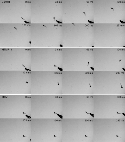
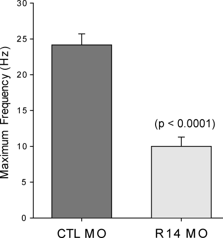
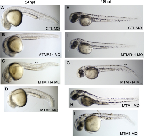
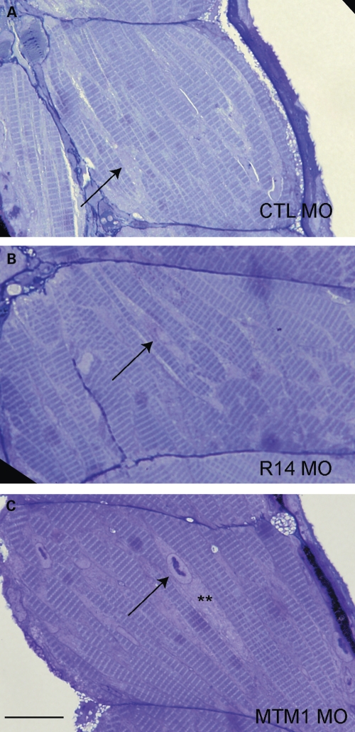
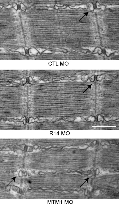
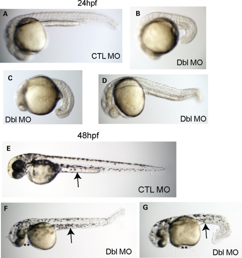
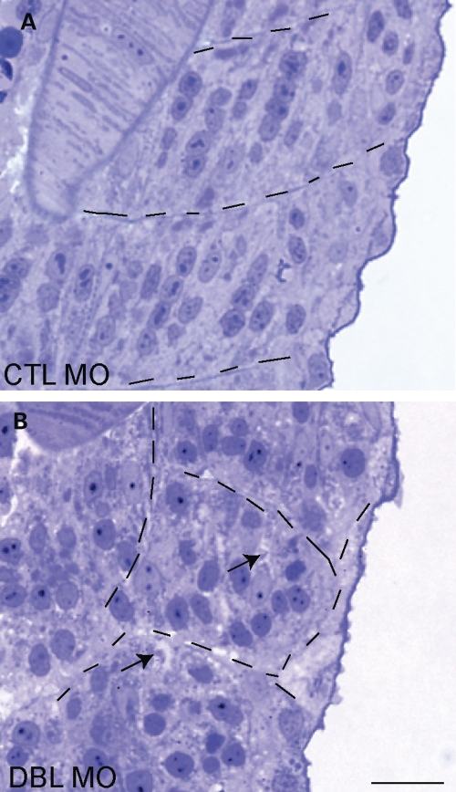
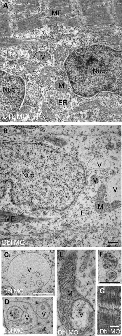
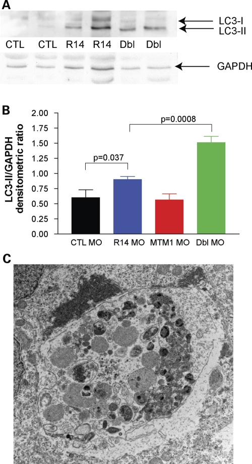
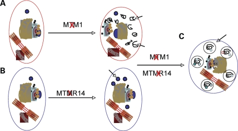
Myotubularins are a family of dual-specificity phosphatases that act to modify phosphoinositides and regulate membrane traffic. Mutations in several myotubularins are associated with human disease. Sequence changes in MTM1 and MTMR14 (also known as Jumpy) have been detected in patients with a severe skeletal myopathy called centronuclear myopathy. MTM1 has been characterized in vitro and in several model systems, while the function of MTMR14 and its specific role in muscle development and disease is much less well understood. We have previously reported that knockdown of zebrafish MTM1 results in significantly impaired motor function and severe histopathologic changes in skeletal muscle that are characteristic of human centronuclear myopathy. In the current study, we examine zebrafish MTMR14 using gene dosage manipulation. As with MTM1 knockdown, morpholino-mediated knockdown of MTMR14 results in morphologic abnormalities, a developmental motor phenotype characterized by diminished spontaneous contractions and abnormal escape response, and impaired excitation-contraction coupling. In contrast to MTM1 knockdown, however, muscle ultrastructure is unaffected. Double knockdown of both MTM1 and MTMR14 significantly impairs motor function and alters skeletal muscle ultrastructure. The combined effect of reducing levels of both MTMR14 and MTM1 is significantly more severe than either knockdown alone, an effect which is likely mediated, at least in part, by increased autophagy. In all, our results suggest that MTMR14 is required for motor function and, in combination with MTM1, is required for myocyte homeostasis and normal embryonic development.


Figures

Figure 1.
Figure 1.
MTMR14 morphants exhibit diminished touch-evoked escape responses. (Top) Tactile stimuli applied to the tail of a wild-type larvae (48–52 hpf) evokes a rapid escape contraction followed by swimming which propels the larvae out of the field of view (n = 27/30). Scale bar 1 mm. (Middle) A similar stimulus applied to the tail of an MTMR14 morphant evokes an escape contraction; however, morphants often failed to generate bouts of swimming sufficient to move out of the field of view (n = 3/30, P = 0.0006). (Bottom) For comparison, an MTM1 morphant performs a reduced escape contraction and low amplitude bending which was insufficient to propel the larvae forward out of the field of view (n = 0/30).
Figure 2.
Figure 2.
MTMR14 morphants fail to respond to high-frequency electrical stimulation. In response to 5 ms depolarizing current injections to +60 mV, skeletal muscle from control embryos routinely contracted above 20 Hz for 10 s (n = 6/6). In contrast, skeletal muscle from MTMR14 morphants failed to contract at 20 Hz (n = 0/6). Values represent the average ± SEM at which muscle could sustain contractions in response to electrical stimuli.
Figure 3.
Figure 3.
MTMR14 morphants exhibit mild morphologic abnormalities. Morphologic appearance at 24 hpf (AD) and 48 hpf (EI) of control morphants (CTL; A and E), MTMR14 morphants (MTMR14 MO; B, C, F, G), and MTM1 morphants (MTM1 MO; D, H, I). MTMR14 morphant appearance ranged from essentially normal (B and F) to exhibiting minor defects in the shape of their midbodies (** in C) or tails (** in G). MTM1 morphants, in comparison, exhibit qualitatively more severe changes in overall appearance, body length and midbody changes.
Figure 4.
Figure 4.
MTMR14 knockdown does not result in obvious histopathologic abnormalities. Skeletal muscle was examined in 72 hpf morphants. Muscle from MTMR14 morphants (R14 MO) was histologically normal and similar to muscle from controls (CTL MO). Arrows identify myonuclei. MTM1 morphants (MTM1 MO), on the other hand, exhibit obvious nuclear (arrow) and perinuclear changes (**). n = 4 per condition. Scale bar = 200 µm.
Figure 5.
Figure 5.
MTMR14 morphant muscle has normal ultrastructure. Ultrastructural analysis of muscle from 48 hpf control (CTL MO), MTMR14 (R14 MO) and MTM1 (MTM1 MO) morphants. Muscle ultrastructure was normal in MTMR14 morphants. In particular, triads were unchanged in appearance (arrow). Conversely, MTM1 morphant muscle has abnormal triads and longitudinal sarcoplasmic reticulum (arrows) (18). n = 4 per condition. Scale bar = 500 nm (34).
Figure 6.
Figure 6.
MTMR14/MTM1 double morphants have severe disturbances in embryonic development. Morphologic analysis of double morphants at 24 hpf (AD) and 48 hpf (EG). When compared with control embryos (CTL MO, A, E), MTMR14/MTM1 double morphants (Dbl MO) exhibited severe alterations in gross morphology. Embryos at 24 hpf were significantly foreshortened with global underdevelopment (B–D). At 48 hpf (F and G), double morphants were small and dysmorphic, with small and bent tails and hypoplastic midbodies. Edema (**) was also present.
Figure 7.
Figure 7.
MTMR14/MTM1 double morphants have abnormal skeletal muscle structure. Light microscopic analysis of semi thin sections from 48 hpf embryos. (A) Control morphants (CTL) had normal developing skeletal muscle. (B) MTMR14/MTM1 double morphants (Dbl MO) had altered skeletal muscle structure, including abnormal appearance and localization of myonuclei and the presence of many small areas that lacked staining (arrows). Myofiber compartments are delineated by hatched lines. Scale bar = 250 µm.
Figure 8.
Figure 8.
Ultrastructural analysis of MTMR14/MTM1 double-morphant skeletal muscle. Electron micrographs of skeletal muscle from 48 hpf embryos. (A and B) Low magnification of MTM1/MTMR14 double morphants (Dbl MO) revealed a paucity of sarcoplasm, decreased endoplasmic reticulum density (ER) and the presence of abnormal vacuolar structures (V) and abnormally appearing mitochondria (M). (CE) Higher magnification examination of the abnormal vacuolar structures, including a vacuole within a mitochondrion (E) and vacuoles with subcellular contents (F). (G) Despite these changes, myofibrils appeared normal. Scale bars = 500 nm.
Figure 9.
Figure 9.
Increased autophagy in MTMR14/MTM1 double morphants. (A) Western blot analysis of protein extracted from 48 hpf embryos. Blots were probed with anti-LC3, stripped and then reprobed with anti-GAPDH. Lanes: 1 and 2, control morphants (CTL); lanes 3 and 4, MTMR14 morphants (R14); and lanes 5 and 6 MTMR14/MTM1 double morphants (Dbl). (B) Quantitation of the ratio of LC3-II levels to GAPDH levels. Average values were as follows: CTL MO = 0.60, R14 MO = 0.90, MTM1 MO = 0.57, Dbl MO = 1.51. P-value of one-way ANOVA = 0.0001. (C) Example of an abnormal autophagic compartment revealed by electron microscopic analysis of a 48 hpf double-morphant embryo. Such structures were observed in three independently analyzed double morphants.
Figure 10.
Figure 10.
Model of subcellular dysfunction with MTM1 and MTMR14 knockdown. (A) Knockdown of MTM1 results in the aberrant accumulation of membranes (arrows) as well as the abnormal appearance of organelles [Fig. 4B and (18)]. (B) Knockdown of MTMR14 causes an induction of autophagy [Fig. 8 and (15)]. (C) We hypothesize that the combination of accumulated autophagic substrates with excessive autophagic induction overwhelms the degradative system. This results in the accumulation of abnormal autophagic vacuoles (arrows) (Fig. 8C) and global cellular dysfunction, as reflected by the unexpectedly severe phenotype of double-morphant embryos.

References

    1. Miaczynska M., Stenmark H. Mechanisms and functions of endocytosis. J. Cell Biol. 2008;180:7–11. doi:10.1083/jcb.200711073. - DOI - PMC - PubMed
    1. Di Paolo G., De Camilli P. Phosphoinositides in cell regulation and membrane dynamics. Nature. 2006;443:651–657. doi:10.1038/nature05185. - DOI - PubMed
    1. McCrea H.J., De Camilli P. Mutations in phosphoinositide metabolizing enzymes and human disease. Physiology (Bethesda) 2009;24:8–16. - PMC - PubMed
    1. Robinson F.L., Dixon J.E. Myotubularin phosphatases: policing 3-phosphoinositides. Trends Cell Biol. 2006;16:403–412. doi:10.1016/j.tcb.2006.06.001. - DOI - PubMed
    1. De Matteis M.A., Godi A. PI-loting membrane traffic. Nat. Cell Biol. 2004;6:487–492. - PubMed

Publication types

MeSH terms

Substances