

A role for the class A penicillin-binding protein PonA2 in the survival of Mycobacterium smegmatis under conditions of nonreplication
- PMID: 20400545
- PMCID: PMC2901687
- DOI: 10.1128/JB.00025-10
A role for the class A penicillin-binding protein PonA2 in the survival of Mycobacterium smegmatis under conditions of nonreplication
Abstract
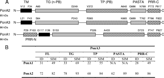
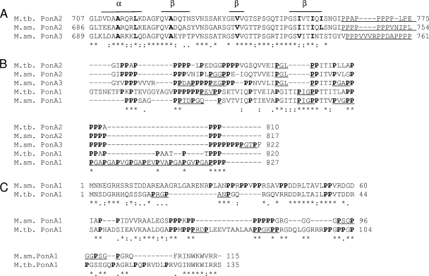
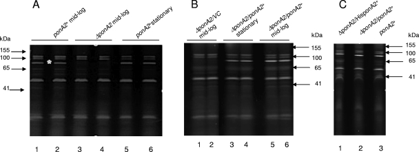
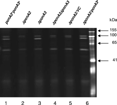
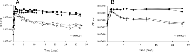
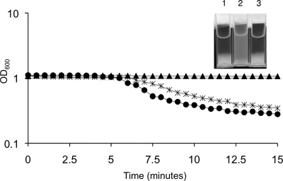
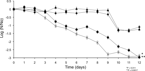
Class A penicillin-binding proteins (PBPs) are large, bifunctional proteins that are responsible for glycan chain assembly and peptide cross-linking of bacterial peptidoglycan. Bacteria in the genus Mycobacterium have been reported to have only two class A PBPs, PonA1 and PonA2, that are encoded in their genomes. We report here that the genomes of Mycobacterium smegmatis and other soil mycobacteria contain an additional gene encoding a third class A penicillin-binding protein, PonA3, which is a paralog of PonA2. Both the PonA2 and PonA3 proteins contain a penicillin-binding protein and serine/threonine protein kinase-associated (PASTA) domain that we propose may be involved in sensing the cell cycle and a C-terminal proline-rich region (PRR) that may have a role in protein-protein or protein-carbohydrate interactions. We show here that an M. smegmatis Delta ponA2 mutant has an unusual antibiotic susceptibility profile, exhibits a spherical morphology and an altered cell surface in stationary phase, and is defective for stationary-phase survival and recovery from anaerobic culture. In contrast, a Delta ponA3 mutant has no discernible phenotype under laboratory conditions. We demonstrate that PonA2 and PonA3 can bind penicillin and that PonA3 can partially substitute for PonA2 when ponA3 is expressed from a constitutive promoter on a multicopy plasmid. Our studies suggest that PonA2 is involved in adaptation to periods of nonreplication in response to starvation or anaerobiosis and that PonA3 may have a similar role. However, the regulation of PonA3 is likely different, suggesting that its importance could be related to stresses encountered in the environmental niches occupied by M. smegmatis and other soil-dwelling mycobacteria.
Figures

Similar articles
-
Structural and binding properties of the PASTA domain of PonA2, a key penicillin binding protein from Mycobacterium tuberculosis.Biopolymers. 2014 Jul;101(7):712-9. doi: 10.1002/bip.22447. Biopolymers. 2014. PMID: 24281824
-
Cell Wall Damage Reveals Spatial Flexibility in Peptidoglycan Synthesis and a Nonredundant Role for RodA in Mycobacteria.J Bacteriol. 2022 Jun 21;204(6):e0054021. doi: 10.1128/jb.00540-21. Epub 2022 May 11. J Bacteriol. 2022. PMID: 35543537 Free PMC article.
-
Loss of a Class A Penicillin-Binding Protein Alters β-Lactam Susceptibilities in Mycobacterium tuberculosis.ACS Infect Dis. 2016 Feb 12;2(2):104-10. doi: 10.1021/acsinfecdis.5b00119. Epub 2015 Dec 21. ACS Infect Dis. 2016. PMID: 27624961
-
Distribution of PASTA domains in penicillin-binding proteins and serine/threonine kinases of Actinobacteria.J Antibiot (Tokyo). 2016 Sep;69(9):660-85. doi: 10.1038/ja.2015.138. Epub 2016 Jan 13. J Antibiot (Tokyo). 2016. PMID: 26758489 Review.
-
Multimodular penicillin-binding proteins: an enigmatic family of orthologs and paralogs.Microbiol Mol Biol Rev. 1998 Dec;62(4):1079-93. doi: 10.1128/MMBR.62.4.1079-1093.1998. Microbiol Mol Biol Rev. 1998. PMID: 9841666 Free PMC article. Review.
Cited by
-
Identification of drivers of mycobacterial resistance to peptidoglycan synthesis inhibitors.Front Microbiol. 2022 Sep 6;13:985871. doi: 10.3389/fmicb.2022.985871. eCollection 2022. Front Microbiol. 2022. PMID: 36147841 Free PMC article.
-
Crystal structures of the transpeptidase domain of the Mycobacterium tuberculosis penicillin-binding protein PonA1 reveal potential mechanisms of antibiotic resistance.FEBS J. 2016 Jun;283(12):2206-18. doi: 10.1111/febs.13738. Epub 2016 May 25. FEBS J. 2016. PMID: 27101811 Free PMC article.
-
Penicillin Binding Proteins and β-Lactamases of Mycobacterium tuberculosis: Reexamination of the Historical Paradigm.mSphere. 2022 Feb 23;7(1):e0003922. doi: 10.1128/msphere.00039-22. Epub 2022 Feb 23. mSphere. 2022. PMID: 35196121 Free PMC article.
-
Mycobacterium tuberculosis sensor kinase DosS modulates the autophagosome in a DosR-independent manner.Commun Biol. 2019 Sep 20;2:349. doi: 10.1038/s42003-019-0594-0. eCollection 2019. Commun Biol. 2019. PMID: 31552302 Free PMC article.
-
The transpeptidase PbpA and noncanonical transglycosylase RodA of Mycobacterium tuberculosis play important roles in regulating bacterial cell lengths.J Biol Chem. 2018 Apr 27;293(17):6497-6516. doi: 10.1074/jbc.M117.811190. Epub 2018 Mar 12. J Biol Chem. 2018. PMID: 29530985 Free PMC article.
References
-
- Ausubel, F. M., R. Brent, R. E. Kingston, D. D. Moore, J. G. Seidman, J. A. Smith, and K. Struhl. 1987. Current protocols in molecular biology. Greene Publishing Associates and Wiley-Interscience, New York, NY.
-
- Azuma, I., D. W. Thomas, A. Adam, J. M. Ghuysen, R. Bonaly, J. F. Petit, and E. Lederer. 1970. Occurrence of N-glycolylmuramic acid in bacterial cell walls. A preliminary survey. Biochim. Biophys. Acta 208:444-451. - PubMed
-
- Basu, J., S. Mahapatra, M. Kundu, S. Mukhopadhyay, M. Nguyen-Disteche, P. Dubois, B. Joris, J. Van Beeumen, S. T. Cole, P. Chakrabarti, and J. M. Ghuysen. 1996. Identification and overexpression in Escherichia coli of a Mycobacterium leprae gene, pon1, encoding a high-molecular-mass class A penicillin-binding protein, PBP1. J. Bacteriol. 178:1707-1711. - PMC - PubMed
Publication types
MeSH terms
Substances
Grants and funding
LinkOut - more resources
Full Text Sources
Molecular Biology Databases
