. 2010 Jun;192(12):3159-73.
doi: 10.1128/JB.00027-10. Epub 2010 Apr 16.

YneA, an SOS-induced inhibitor of cell division in Bacillus subtilis, is regulated posttranslationally and requires the transmembrane region for activity

Affiliations

YneA, an SOS-induced inhibitor of cell division in Bacillus subtilis, is regulated posttranslationally and requires the transmembrane region for activity

Allison H Mo et al. J Bacteriol. 2010 Jun.

Abstract

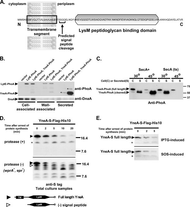
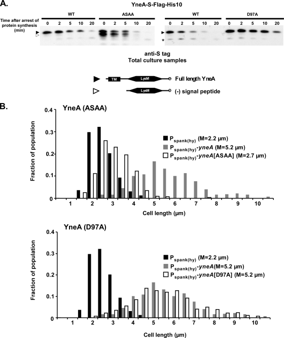
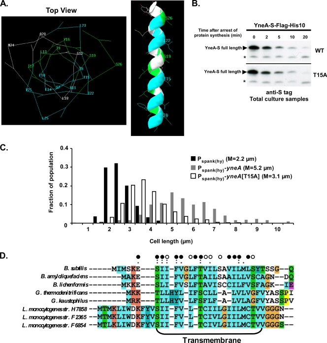
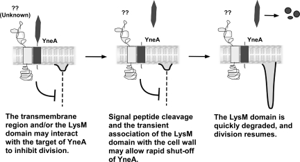
Cell viability depends on the stable transmission of genetic information to each successive generation. Therefore, in the event of intrinsic or extrinsic DNA damage, it is important that cell division be delayed until DNA repair has been completed. In Bacillus subtilis, this is accomplished in part by YneA, an inhibitor of division that is induced as part of the SOS response. We sought to gain insight into the mechanism by which YneA blocks cell division and the processes involved in shutting off YneA activity. Our data suggest that YneA is able to inhibit daughter cell separation as well as septum formation. YneA contains a LysM peptidoglycan binding domain and is predicted to be exported. We established that the YneA signal peptide is rapidly cleaved, resulting in secretion of YneA into the medium. Mutations within YneA affect both the rate of signal sequence cleavage and the activity of YneA. YneA does not stably associate with the cell wall and is rapidly degraded by extracellular proteases. Based on these results, we hypothesize that exported YneA is active prior to signal peptide cleavage and that proteolysis contributes to the inactivation of YneA. Finally, we identified mutations in the transmembrane segment of YneA that abolish the ability of YneA to inhibit cell division, while having little or no effect on YneA export or stability. These data suggest that protein-protein interactions mediated by the transmembrane region may be required for YneA activity.


Figures

FIG. 1.
FIG. 1.
YneA delays cell division. (A) Strains overexpressing yneA are unable to form single colonies on plates. AM69 [amyE::Pspank(hy)-yneA] and AM70 [amyE::Pspank(hy)] were streaked onto LB agar containing 1 mM IPTG and grown overnight at 30°C. (B) Cells overexpressing yneA are elongated. AM62 (amyE::cat) and AM93 [amyE::Pspank(hy)-yneA] were induced with 1 mM IPTG as described in Materials and Methods. One hour after induction, cells were stained with FM4-64 and DAPI and then visualized. Bars, 5 μm. (C) Cell length distributions from the same experiment as that for panel B. At least 300 cells were measured for each strain. The experiment was repeated more than four times under essentially the same conditions. The graph shown here is from a representative experiment. (D) Overexpression of yneA delays the first cell division during spore outgrowth. Spore preparations of AM69 [amyE::Pspank(hy)-yneA] and AM70 [amyE::Pspank(hy)] were resuspended in GMD medium containing 1 mM IPTG and incubated at 37°C. Samples were taken at the indicated times. Cell membranes were stained with FM4-64. Cells or cell chains with one or more visible septa were scored as having divided. At least 125 germinated cells were counted for each sample.
FIG. 2.
FIG. 2.
YneA delays FtsZ ring formation. (A) FtsZ-YFP localization in cells expressing yneA. AM293 [amyE::Pxyl-ftsZ-yfp thrC::Pspank(hy)-yneA] was induced with 1 mM IPTG (+YneA) and 0.5% xylose. IPTG was omitted in one culture (−YneA). One hour after induction, cells were stained with FM4-64 and visualized. Bars, 1 μm. (B) YneA delays FtsZ ring assembly. Cell lengths were measured as described for Fig. 1C and were plotted with the length on the x axis and the fraction of the population at each length on the left y axis (bars). Fluorescent bands seen in the YFP channel that were not associated with completed septa or cell poles were scored as nonpolar FtsZ rings. The average number of these FtsZ rings per cell at each length was plotted on the right y axis (lines). More than 300 cells from each sample were measured. Of the >200 total FtsZ rings counted in each sample, between 15% (−YneA) and 37% (+YneA) were nonpolar. This experiment is representative of two independent trials.
FIG. 3.
FIG. 3.
YneA inhibits cell separation. (A) Cells overexpressing yneA remain in chains after the onset of stationary phase. Images are from the same experiment as that described for Fig. 1C. Cells were stained with FM4-64 and DAPI and were visualized 5 h after induction of yneA with 1 mM IPTG (stationary phase). Bars, 5 μm. (B) Quantitation of cell chain lengths from the same experiment as that for panel A. Samples were taken 5 h after induction of yneA. The number of cells per chain was determined by comparing phase-contrast and FM4-64-stained images. At least 400 cells (>200 chains) were counted for each strain. This experiment is representative of two independent trials under essentially the same conditions. (C) AM62 (amyE::cat) and AM93 [amyE::Pspank(hy)-yneA] were grown overnight on LB agar, resuspended in T-base, diluted to an OD600 of ∼0.02 in CH (rich) medium, and grown at 37°C to exponential growth phase (OD600 = 0.3 to 0.5). Cells were then resuspended in SM (minimal) medium containing 1 mM IPTG. Samples were taken at the time of resuspension (top graph) and 90 min after resuspension (bottom graph), and chain lengths were quantified as in panel B. This experiment is representative of two independent trials.
FIG. 4.
FIG. 4.
Division resumes rapidly after yneA expression is shut off. AM69 [amyE::Pspank(hy)-yneA] was induced with 1 mM IPTG. IPTG was omitted in one culture throughout the experiment, as a control (−IPTG; white bars). One hour after induction, cells were centrifuged and resuspended in an equal volume of LB, with (+IPTG; black bars) or without (+IPTG, −IPTG; gray bars) 1 mM IPTG. Samples were taken 0, 20, 40, and 60 min after resuspension. Cells were stained and measured as described in the legend to Fig. 1C. This experiment is representative of two independent trials under essentially the same conditions.
FIG. 5.
FIG. 5.
YneA is rapidly cleaved and secreted into the medium. (A) Predicted domains of YneA. Predictions are based on sequence analysis using SignalP and Pfam. (B) YneA is secreted into the medium and does not associate strongly with the cell wall. Asterisks (*) indicate probable LytE-PhoA degradation products. AM206 [thrC::Pspank(hy)], AM218 (lytE::lytE-phoA), and AM222 [lytE::lytE-phoA thrC::Pspank(hy)-yneA-phoA] were grown in GYE minimal medium and induced with 1 mM IPTG. Two hours after induction, cultures were centrifuged and then separated into cell, wall, and secreted fractions as described in Materials and Methods. Samples were separated in a 10% SDS-polyacrylamide gel. Each lane contains 2 μg of cell fraction, 0.5 μg of wall fraction, or 0.5 μg of secreted fraction. Immunoblots were performed with either anti-PhoA antibody or anti-DnaA antibody. (C) YneA export is SecA dependent. AM98 [thrC::Pspank(hy)-yneA-phoA] and AM280 [secA314(Ts) thrC::Pspank(hy)-yneA-phoA] were incubated at either 30°C or 45°C in GYE medium containing 1 mM IPTG. After 1 h of induction, cultures were centrifuged and the secreted fraction prepared as described in Materials and Methods. Samples were separated in a 10% SDS-polyacrylamide gel. Each lane contains 2 μg of cell fraction or ∼0.5 μg of secreted fraction. Immunoblots were performed with anti-PhoA antibody. (D) Full-length YneA is rapidly cleaved. Black arrowheads, full-length YneA-S-Flag-His10 (∼16.9 kDa); open arrowheads, C-terminal portion of YneA-S-Flag-His10 lacking signal peptide (∼13.5 kDa); asterisk, nonspecific band. AM199 [thrC::Pspank(hy)-yneA-S-flag-his10] and AM200 [epr::tet wprA::kan thrC::Pspank(hy)-yneA-S-flag-his10] were induced with 1 mM IPTG. After 1 h of induction, protein synthesis was inhibited with 200 μg ml−1 chloramphenicol, and whole culture samples were removed at the indicated times after drug addition. S-tagged proteins were isolated as described in Materials and Methods and separated in a 13.8% SDS-polyacrylamide gel. Immunoblots were performed using diluted (1:5,000) anti-S primary antibody. (E) Induction of the SOS response does not affect the stability of YneA. AM199 [thrC::Pspank(hy)-yneA-S-flag-his10] and AM214 [yneA::Pspank(hy)-yneA-S-flag-his10] were induced with 1 mM IPTG and 1 μg ml−1 mitomycin C, respectively. After 1 h of induction, protein synthesis was arrested, and samples were taken and treated as described for panel D.
FIG. 6.
FIG. 6.
Full-length YneA may be the active form. (A) ASAA and D97A mutations affect the stability of full-length and cleaved YneA. Black arrowheads, full-length YneA-S-Flag-His10; open arrowheads, C-terminal portion of YneA-S-Flag-His10 lacking the signal peptide; asterisk, nonspecific band. AM199 [thrC::Pspank(hy)-yneA-S-flag-his10], AM202 [thrC::Pspank(hy)-yneA(ASAA)-S-flag-his10], and AM228 [thrC::Pspank(hy)-yneA(D97A)-S-flag-his10] were grown and samples were prepared as described in the legend to Fig. 5D. Proteins were separated in a 13.8% SDS-polyacrylamide gel, and immunoblots were performed using diluted (1:5,000) anti-S primary antibody. (B) YneA(ASAA) is inactive, while YneA(D97A) remains active. AM69 [amyE::Pspank(hy)-yneA], AM70 [amyE::Pspank(hy)], AM133 [amyE::Pspank(hy)-yneA(ASAA)], and AM155 [amyE::Pspank(hy)-yneA(D97A)] were grown and induced, and cells were stained and measured as described in the legend to Fig. 1C. This experiment is representative of two independent trials.
FIG. 7.
FIG. 7.
Mutations in the transmembrane region inactivate YneA. (A) Mutations that inactivate YneA cluster on one face of the predicted transmembrane region. The transmembrane region was modeled as an alpha-helix, using Swiss-PDP Viewer. Residues are colored as follows: blue indicates alanine substitutions that inactivate YneA, green indicates alanine substitutions that have little or no effect on YneA, and white indicates residues that were not mutated or tested. (B) The T15A mutation does not affect the stability of YneA. AM199 [thrC::Pspank(hy)-yneA-S-flag-his10] and AM261 [thrC::Pspank(hy)-yneA(T15A)-S-flag-his10] were grown and samples were prepared as described in the legend to Fig. 5D. Proteins were separated in a 13.8% SDS-polyacrylamide gel, and immunoblots were performed using diluted (1:5,000) anti-S primary antibody. Asterisks indicate nonspecific bands. (C) The T15A mutation reduces YneA function. AM69 [amyE::Pspank(hy)-yneA], AM70 [amyE::Pspank(hy)], and AM239 [amyE::Pspank(hy)-yneA(T15A)] were grown and induced (in the same experiment as that for Fig. 6B), and cells were stained and measured as described in the legend to Fig. 1C. This experiment is representative of two independent trials. (D) Many inactivating mutations are in conserved residues. Alignment of transmembrane domains in YneA orthologues was performed using ClustalX2.0. Open circles, alanine substitutions with little or no effect on YneA activity; closed circles, alanine substitutions that inactivate YneA; single and double dots indicate conserved amino acid groups.
FIG. 8.
FIG. 8.
Proposed model for YneA activity.

Similar articles

Cited by

References

    1. Antelmann, H., H. Yamamoto, J. Sekiguchi, and M. Hecker. 2002. Stabilization of cell wall proteins in Bacillus subtilis: a proteomic approach. Proteomics 2:591-602. - PubMed
    1. Au, N., E. Kuester-Schoeck, V. Mandava, L. E. Bothwell, S. P. Canny, K. Chachu, S. A. Colavito, S. N. Fuller, E. S. Groban, L. A. Hensley, T. C. O'Brien, A. Shah, J. T. Tierney, L. L. Tomm, T. M. O'Gara, A. I. Goranov, A. D. Grossman, and C. M. Lovett. 2005. Genetic composition of the Bacillus subtilis SOS system. J. Bacteriol. 187:7655-7666. - PMC - PubMed
    1. Autret, S., A. Levine, I. B. Holland, and S. J. Seror. 1997. Cell cycle checkpoints in bacteria. Biochimie 79:549-554. - PubMed
    1. Bateman, A., and M. Bycroft. 2000. The structure of a LysM domain from E. coli membrane-bound lytic murein transglycosylase D (MltD). J. Mol. Biol. 299:1113-1119. - PubMed
    1. Bendtsen, J. D., H. Nielsen, G. von Heijne, and S. Brunak. 2004. Improved prediction of signal peptides: SignalP 3.0. J. Mol. Biol. 340:783-795. - PubMed

Publication types

MeSH terms

LinkOut - more resources