. 2010 Jul;88(1):169-80.
doi: 10.1189/jlb.0809548. Epub 2010 Apr 16.

The myeloid 7/4-antigen defines recently generated inflammatory macrophages and is synonymous with Ly-6B

Affiliations

The myeloid 7/4-antigen defines recently generated inflammatory macrophages and is synonymous with Ly-6B

Marcela Rosas et al. J Leukoc Biol. 2010 Jul.

Erratum in

  • J Leukoc Biol. 2011 Nov;90(5):1035
  • Erratum.
    [No authors listed] [No authors listed] J Leukoc Biol. 2011 Nov;90(5):1035. doi: 10.1189/jlb.0809548err. J Leukoc Biol. 2011. PMID: 29360164 Free PMC article. No abstract available.

Abstract

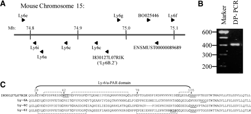
This study aimed to identify the inflammation-associated 7/4-antigen, which is highly expressed on neutrophils, inflammatory monocytes, some activated macrophages, as well as on bone marrow myeloid-restricted progenitors. The high expression on inflammatory cells is suggestive of a role in inflammation and makes the 7/4-antigen a potential target for the manipulation of inflammatory cells. Consistent with this, the 7/4-antibody mediates specific depletion of 7/4-expressing neutrophils and monocytes. We have identified the 7/4-antigen as a 25- to 30-kDa GPI-anchored glycoprotein synonymous with the Ly-6B.2 alloantigen. We characterized the expression of Ly-6B during the inflammatory reaction induced by zymosan. During the later stages of an experimental, acute, self-resolving inflammatory response, we found that Ly-6B is differentially expressed on macrophages. Ly-6B-expressing macrophages also express more MHCII, CIITA, CCR2, Ly-6C, and CD62L than the Ly-6B-negative macrophages, which in turn, express more of the resident tissue macrophage marker SIGN-R1 and higher CD11b and F4/80. Ly-6B-expressing macrophages incorporate more BrdU than their Ly-6B-negative contemporaries when fed during the resolution phase of the acute inflammatory response. Thus, Ly-6B expression on mature macrophages defines a subset of recently generated inflammatory macrophages that retain monocytic markers and is hence a surrogate marker of macrophage turnover in inflammatory lesions. The definition of the 7/4:Ly-6B antigen will allow further characterization and specific modulation of Ly-6B-expressing cells in vivo.


Figures

Figure 1.
Figure 1.
Distribution of the 7/4-antigen. (A) Different tissues were obtained and pooled from two C57BL/6 mice and separated by SDS-PAGE. Western blotting with the 7/4-antibody was performed as indicated in Materials and Methods. (B) Peripheral blood leukocytes (129) were labeled with CD11b, F4/80, Ly-6C, and 7/4, as indicated previously. Monocytes were first identified on the FSC/SSC plot and then as CD11b+F4/80+ cells, as described previously [20]. Expression of the 7/4-antigen determined two populations of monocytes (7/4+ and 7/4), which are comparable with the populations established by the expression of Ly-6C (Ly-6C+and Ly-6C). Data are derived from blood pooled from four independent animals, and similar results were obtained from three independent experiments with the Gr-1 antibody. (C) Depletion of peripheral myeloid cells by i.p. administration of the 7/4-antibody. Compared with isotype control-injected animals (open symbols), 7/4-injected mice (closed symbols) had substantially reduced numbers of neutrophils (Neu; identified as SSChiCD11b+Ly-6GhiGr-1hi) and Gr-1+ monocytes (Mo; SSClowF4/80+CD11b+Gr-1+) in the periphery. Gr-1 monocytes and eosinophils (Eos; SSCv.hiCD11b+F4/80+Gr-1int) are also shown. Data are expressed as the percentage of peripheral blood leukocytes defined as above, normalized to percentage present in isotype control-injected mice. Each symbol represents a single mouse, and horizontal bars denote means. Data are derived from the larger of two independent experiments and were analyzed by two-way ANOVA with Bonferonni post-tests (*, P<0.05; ***, P<0.001).
Figure 2.
Figure 2.
7/4-Antigen is a low molecular weight N-glycosylated polymorphic protein with a GPI anchor. (A) Bone marrow lysates from C57BL/6 and Balb/c mice were separated by SDS-PAGE under nonreducing and reducing conditions. Western blotting with the 7/4-antibody was performed as indicated above. (B) Subcellular fractionation of the nitrogen-cavitated, thioglycollate-elicited, enriched neutrophil population resulted in the separation of membrane, cytoplasm, and nucleus fractions. Each fraction, as well as total lysate, was separated by SDS-PAGE, followed by Western blotting with the 7/4-antibody or Gr-1 as indicated previously. (C) Thioglycollate-elicited, neutrophil-rich membranes obtained from subcellular fractionation were treated with or without PNGase and Western blotted with the 7/4-antibody. (D) Monocytes and neutrophils were treated with PI-PLC at 4°C or 37°C and then labeled with 7/4, Gr-1, or isotype control antibodies for flow cytometric analysis. All experiments were conducted at least twice.
Figure 3.
Figure 3.
7/4-Antigen is genetically linked to the chromosome 15 Ly-6 family. (A) A congenic Balb/c.7/4+ mouse generated by six generations of backcrossing exhibited expression of the 7/4-antigen on neutrophils in peripheral blood, and this was compared with the expression of Ly-6A.2 on lymphocytes of C57BL/6, Balb/c, and the congenic 7/4+ mouse. Staining in the Balb/c.7/4+ mice is representative of six independent animals. (B) Flow cytometric analysis to assess the expression of the 7/4-antigen on neutrophils derived from C57BL/6 and NZB mice. (C) Spleen lysates from various mouse strains including C57BL/6, 129, NZB, CBA, and Balb/c were separated by SDS-PAGE and Western blotted with the 7/4-antibody. Analysis of NZB mice is representative of two independent animals.
Figure 4.
Figure 4.
Genomic location of the Ly-6-family members and protein alignment. (A) Distribution of the Ly-6 genes on the chromosome 15 according to ENSEMBL. A new identified member called I830127L07Rik is located between the Ly6g and Ly6c genes. (B) A single degenerate RT-PCR product of the expected size was obtained after isolating RNA from inflammatory-peritoneal neutrophils and amplification of Ly-6-related, expressed sequences using degenerate primers (DP). (C) Comparison of the primary amino acid sequence of I830127L07Rik to Ly-6A, Ly-6C, and Ly-6I. All of these proteins contain the Ly-6/u-PAR domain with 10 conserved cysteines (bold) linked by disulfide bonds (indicated by dashed brackets). N-Glycosylation was assessed by NetNGlyc, and NXS/T sequons were found in I830127L07Rik and Ly-6I (bold and underlined). Prediction of GPI anchor sites was performed with big PI predictor [23], and these sites are underlined. mb, Megabase.
Figure 5.
Figure 5.
7/4-Antigen is synonymous with Ly-6B.2. (A) NIH3T3-stable cell lines expressing the indicated Ly-6 proteins were analyzed by Western blot with the 7/4-antibody, which only recognizes the protein encoded by the complete I830127L07Rik sequence. (B) RAW264.7 cells expressing the I830127L07Rik sequence were labeled with the 7/4-antibody or SK38.86 ascites (black line) and empty vector-transduced control (filled histogram). Analysis was performed by flow cytometry. Data confirming flow cytometric recognition of I830127L07Rik by the 7/4-antibody was obtained twice with RAW264.7 cells and once with NIH3T3 cells. (C) NIH3T3 expressing Ly-6B.2 were cultured on coverslips and stained with FITC-labeled 7/4-antibody or IgG2a (isotype control). Visualization of Ly-6B.2 at the plasma membrane was done by fluorescence microscopy. Similar results were obtained in a second experiment using independently generated, retrovirally transduced Ba/F3 cells (not shown). (D) Schematic representation of the three exons coding for Ly-6B. The third exon on the sequence of Balb/c mice (Ly-6.1) holds a premature stop codon.
Figure 6.
Figure 6.
Expression of Ly-6B during zymosan-induced peritoneal inflammation. (A) C57BL/6 mice were injected with 2 × 10⁷ zymosan particles, and peritoneal lavages were collected after 4, 18, 72, and 168 h. Cells were stained with F4/80, CD11b, and Ly-6B as indicated previously. The first panel shows the expression of F4/80 and CD11b at different time-points. At 4 h, residual resident macrophages (F4/80highCD11bhigh) and inflammatory monocytes (F4/80lowCD11blow) are readily identified. By 18 h, resident macrophages (F4/80highCD11bhigh) and inflammatory monocyte/macrophage (F4/80lowCD11blow) are evident. The F4/80 and CD11b profiles of these two populations “merge” during the 72- and 168-h resolution phases, and they are separated arbitrarily into three populations. The additional population with intermediate expression of F4/80 and CD11b (F4/80intCD11bint) emerges at 72 h and remains present at 168 h. Eosinophils (E) and neutrophils (N) are identified as indicated. Based on the F4/80:CD11bhigh,int,low gates, the second row of panels illustrates the changes of Ly-6B expression through the inflammatory response compared with F4/80. The novel Ly-6B+F4/80int “resolution phase” population is indicated. The third row of panels displays the expression of F4/80 associated with size of the cells (FSC area). (B) The expression of Ly-6B on the cells within the arbitrary F4/80highCD11bhigh, F4/80intCD11bint, and F4/80lowCD11blow gates indicated in A is represented as the mean ± sem of absolute cell counts from independent animals (three mice/group). Total counts for each population are shown with those that are Ly-6B+, indicated with shaded areas of bars. These data are consistent with prior publications at 4 and 18 h [4, 5, 7] and representative of two independent repeats, and the separation of macrophages into Ly-6B+ and Ly-6B subsets at Days 3 and 7 is representative of two and seven independent experiments, respectively.
Figure 7.
Figure 7.
Ly-6B and Ly-6B+ cells display different phenotypes. C57BL/6 mice were injected with 2 × 10⁷ zymosan particles, and peritoneal lavages were collected after 7 days (168 h). Cells were stained with the indicated markers and analyzed by flow cytometry. Data are compiled from representative plots obtained with three independent mice or pooled cells from three mice from two independent experiments. (B) Cells were sorted by MoFlo for RNA isolation, as indicated in Materials and Methods. Plots are representative of three independent cell-sorting experiments with cells pooled from C57BL/6 mice. (C) Semi-quantitative RT-PCR analysis of select transcripts in cells sorted based on Ly-6B expression, as described in Materials and Methods. Similar differences were observed in independent RNA analysis of three separately sorted pairs of samples.
Figure 8.
Figure 8.
Proliferation of macrophages assessed by BrdU incorporation. C57BL/6 mice were injected with 2 × 10⁷ zymosan particles. Mice were fed with 0.8 mg/ml BrdU in drinking water for 2 and 4 days before the 7-day time-point. A group of mice without BrdU feeding was used as control. Peritoneal lavages were collected after 7 days, and BrdU detection was performed using the BrdU flow kit (BD PharMingen), as indicated in Materials and Methods. (A) Representative flow cytometric analysis of mice showing the incorporation of BrdU after 2 and 4 days by Ly-6B and Ly-6B+ cells. Gating was determined using mice that had not been fed BrdU. (B) Percentage of BrdU incorporation by Ly-6B and Ly-6B+ cells. Each symbol represents an individual mouse from the same experiment respresented in A, and background data, from mice that had not been fed BrdU, were subtracted. Data are representative of one and two (2 days) independent experiments, and contemporary experiments were analyzed by two-way ANOVA as indicated. Dashed lines indicate the samples derived from the same mice.

References

    1. Hirsch S, Gordon S. Polymorphic expression of a neutrophil differentiation antigen revealed by monoclonal antibody 7/4. Immunogenetics. 1983;18:229–239. - PubMed
    1. Gordon S, Keshav S, Stein M. BCG-induced granuloma formation in murine tissues. Immunobiology. 1994;191:369–377. - PubMed
    1. Lee S H, Crocker P, Gordon S. Macrophage plasma membrane and secretory properties in murine malaria. Effects of Plasmodium yoelii blood-stage infection on macrophages in liver, spleen, and blood. J Exp Med. 1986;163:54–74. - PMC - PubMed
    1. Henderson R B, Hobbs J A, Mathies M, Hogg N. Rapid recruitment of inflammatory monocytes is independent of neutrophil migration. Blood. 2003;102:328–335. - PubMed
    1. Taylor P R, Brown G D, Geldhof A B, Martinez-Pomares L, Gordon S. Pattern recognition receptors and differentiation antigens define murine myeloid cell heterogeneity ex vivo. Eur J Immunol. 2003;33:2090–2097. - PubMed

Publication types