. 2010 Aug;89(8):785-90.
doi: 10.1177/0022034510366903. Epub 2010 Apr 16.

Cleavage site specificity of MMP-20 for secretory-stage ameloblastin

Affiliations

Cleavage site specificity of MMP-20 for secretory-stage ameloblastin

Y-H P Chun et al. J Dent Res. 2010 Aug.

Abstract

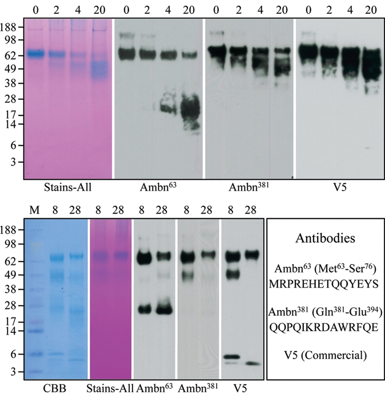
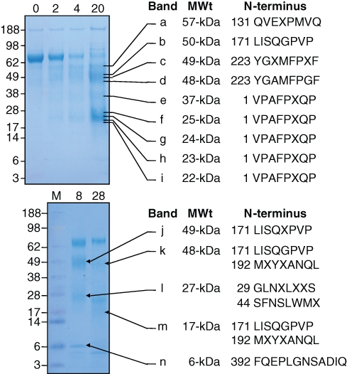
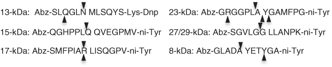
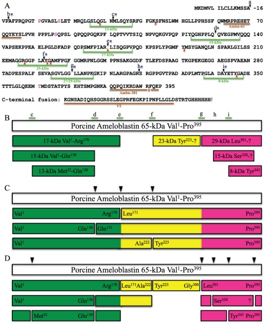
Ameloblastin is processed by protease(s) during enamel formation. We tested the hypothesis that MMP-20 (enamelysin) catalyzes the cleavages that generate secretory-stage ameloblastin cleavage products. We isolated a 23-kDa ameloblastin cleavage product from developing enamel and determined its N-terminus sequence. Ameloblastin was stably expressed and secreted from HEK293-H cells, purified, and digested with MMP-20 or Klk4 (kallikrein 4). The digests were analyzed by SDS-PAGE and Western blotting, and cleavage products were characterized by N-terminal sequencing. Six fluorescent peptides were digested with MMP-20 and Klk4 and analyzed by RP-HPLC and by mass spectrometry. MMP-20 cleaved each peptide exactly at the sites corresponding to ameloblastin cleavages catalyzed in vivo. Klk4 cleaved ameloblastin and the fluorescent peptides at sites not observed in vivo, and cleaved at only a single correct site: before Leu(171). We conclude that MMP-20 is the enzyme that processes ameloblastin during the secretory stage of amelogenesis, and we present a hypothesis about the sequence of ameloblastin cleavages.


Conflict of interest statement

All authors declare that there are no conflicting interests.

Figures

Figure 1.
Figure 1.
Digestion of rAmbn with rMMP-20 and Klk4. (Top) The rAmbn was digested by MMP-20 for 0, 2, 4, and 20 hrs. From left to right: Samples from these digests were separated by SDS-PAGE and stained with Stains-all, or transferred to a membrane for Western blotting with the primary antibodies Ambn63, Ambn381, or the V5 antibody, which recognizes the extreme C-terminus of the ameloblastin fusion protein. Note that the Ambn63 antibody immunostained the intact protein at ~ 65-kDa and a smear of bands ranging from ~13- to 28-kDa, while the Ambn381 and V5 antibodies recognized the intact protein and a smear of bands ranging from ~45- to 60-kDa. This shows that MMP-20 cleaved rAmbn at multiple sites on the N-terminal side of the protein. (Bottom) The rAmbn was digested by Klk4 for 8 and 28 hrs. From left to right: Samples from these digests were separated by SDS-PAGE and stained with CBB or Stains-all, or transferred to a membrane for Western blotting with the primary antibodies Ambn63, Ambn381, or the V5 antibody. The peptides used to synthesize the Ambn63 and Ambn381 are shown at the right.
Figure 2.
Figure 2.
N-terminal sequencing of rAmbn digestion products. (Top) The rAmbn was digested by MMP-20 for 0, 2, 4, and 20 hrs, and samples from these digests were separated by SDS-PAGE and stained with CBB, or transferred to a membrane, which was lightly stained with CBB, and 9 bands (a through i) were excised. The apparent molecular weight and N-terminal sequence of each band are indicated. Bands f, h, and i contained the sequences indicated, starting with Val1, as well as sequences starting with Ala3. It is not known if MMP-20 makes the cleavage before Ala3 or if the signal peptidase alternatively cleaves after Val1 and Ala3. (Bottom) The rAmbn was digested by Klk4 for 8 and 28 hrs, and samples from these digests were separated by SDS-PAGE and stained with CBB, or transferred to a membrane, which was lightly stained with CBB, and 5 bands (j through n) were excised. The N-terminal sequences of the bands are indicated. Bands k, l, and m gave 2 sequences.
Figure 3.
Figure 3.
FRET peptide cleavage sites identified by mass spectrometry. Arrowheads above each peptide sequence indicate sites cleaved by MMP-20. Arrowheads below each peptide sequence indicate sites cleaved by Klk4. MMP-20 cleaved each peptide exactly at the sites corresponding to ameloblastin cleavages catalyzed in vivo: on the N-terminal sides of Met32, Gln131, Leu171, Tyr223, Leu301, and Tyr343.
Figure 4.
Figure 4.
Ambn395 sequence and cleavage sites. (A) Sites where ameloblastin is cleaved in vivo are lettered: (a) Val1, signal peptidase cleavage site (Uchida et al., 1995); (b) Ala3, possible alternative signal peptide cleavage site or MMP-20 site (Iwata et al., 2007); (c) Met31, N-terminus of the 13-kDa (Hu et al., 1997); (d) Gln131, C-terminus of the 13- and 15-kDa (Hu et al., 1997); (e) Leu171, C-terminus of the 17-kDa (Fukae et al., 2006); (f) Tyr223 (this report); (g) Leu301, N-terminus of 27- and 29-kDa calcium-binding proteins (Murakami et al., 1997); (h) N-terminus of the 13- and 15-kDa calcium-binding proteins (Yamakoshi et al., 2001); and (i) N-terminus of an 8-kDa peptide (Yamakoshi et al., 2006a). Green lines indicate synthetic peptides used in this study. Blue dots indicate sites shown to be cleaved by MMP-20 in vitro. Red arrowheads indicate sites shown to be cleaved by Klk4 in vitro. Brown lines indicate antigen-binding sites for antibodies used in this study. (B) Porcine ameloblastin cleavage products that have been isolated and characterized in vivo. White bar shows the porcine ameloblastin 65-kDa protein; colored bars are cleavage products. Colors in bars indicate the N-terminal region (Val1 to Arg170; green), middle (Leu171 to Gly300; yellow), and C-terminal region (Leu301 to Pro395; mauve). (C) Initial cleavage products based upon the digestion of rAmbn by MMP-20 in vitro. Arrowheads indicate initial cleavage sites. (D) Secondary MMP-20 cleavages (arrowheads) continue the processing of ameloblastin to yield additional cleavage products. Labeled ends of bars have been confirmed experimentally; unlabeled bars are generated by multiple MMP-20 cleavages, but have not been confirmed experimentally.

References

    1. Bartlett JD, Simmer JP, Xue J, Margolis HC, Moreno EC. (1996). Molecular cloning and mRNA tissue distribution of a novel matrix metalloproteinase isolated from porcine enamel organ. Gene 183:123-128 - PubMed
    1. Bartlett JD, Ryu OH, Xue J, Simmer JP, Margolis HC. (1998). Enamelysin mRNA displays a developmentally defined pattern of expression and encodes a protein which degrades amelogenin. Connect Tissue Res 39:101-109 - PubMed
    1. Beyeler M, Schild C, Lutz R, Chiquet M, Trueb B. (2010). Identification of a fibronectin interaction site in the extracellular matrix protein ameloblastin. Exp Cell Res [Epub ahead of print], January 4, 2010 (in press) - PubMed
    1. Brunati AM, Marin O, Bisinella A, Salviati A, Pinna LA. (2000). Novel consensus sequence for the Golgi apparatus casein kinase, revealed using proline-rich protein-1 (PRP1)-derived peptide substrates. Biochem J 351(Pt 3):765-768 - PMC - PubMed
    1. Caterina JJ, Skobe Z, Shi J, Ding Y, Simmer JP, Birkedal-Hansen H, et al. (2002). Enamelysin (matrix metalloproteinase 20)-deficient mice display an amelogenesis imperfecta phenotype. J Biol Chem 277:49598-49604 - PubMed

Publication types

LinkOut - more resources