

Computational fluid dynamics analysis of thrombosis potential in left ventricular assist device drainage cannulae
- PMID: 20400890
- PMCID: PMC2875940
- DOI: 10.1097/MAT.0b013e3181d861f1
Computational fluid dynamics analysis of thrombosis potential in left ventricular assist device drainage cannulae
Abstract
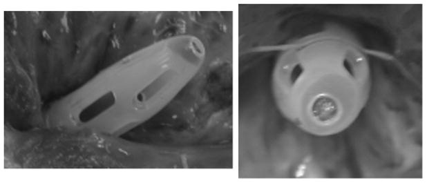
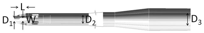
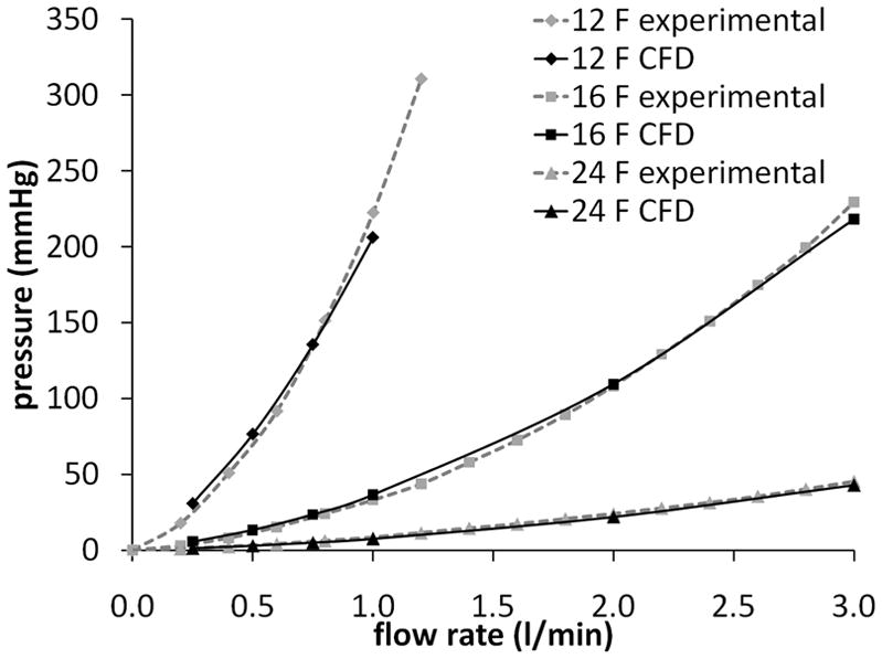
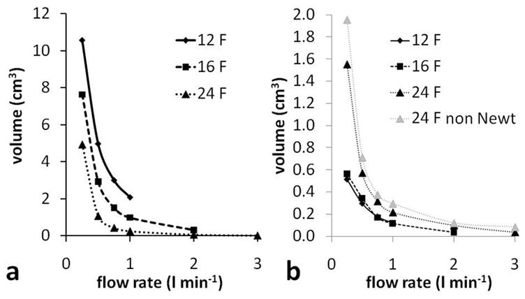
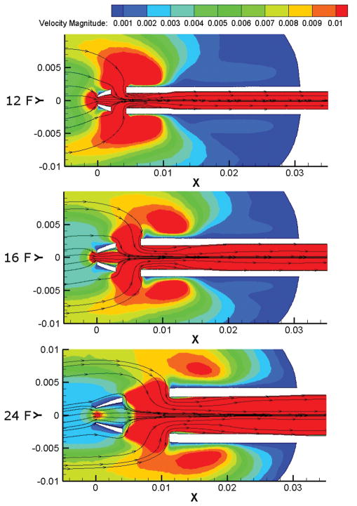
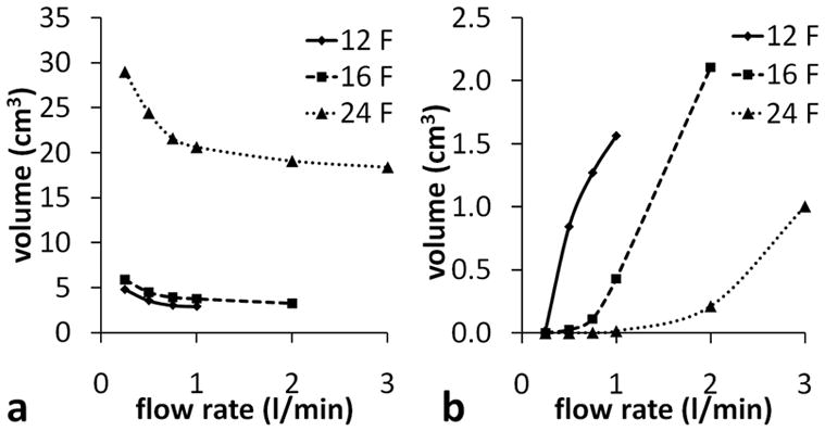
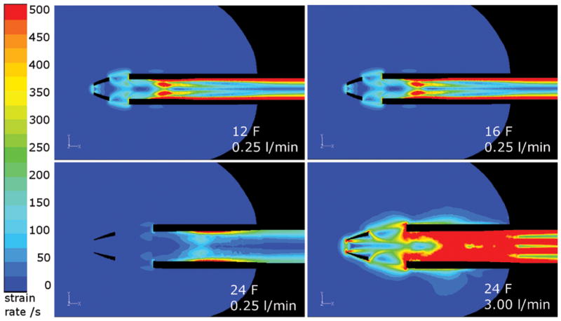
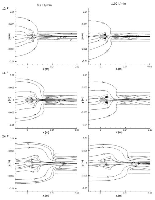
Cannulation is necessary when blood is removed from the body, for example in hemodialysis, cardiopulmonary bypass, blood oxygenators, and ventricular assist devices. Artificial blood contacting surfaces are prone to thrombosis, especially in the presence of stagnant or recirculating flow. In this work, computational fluid dynamics was used to investigate the blood flow fields in three clinically available cannulae (Medtronic DLP 12, 16, and 24 F), used as drainage for pediatric circulatory support and to calculate parameters that may be indicative of thrombosis potential. The results show that using the 24 F cannula below flow rates of about 0.75 L/min produces hemodynamic conditions, which may increase the risk of blood clotting within the cannula. No reasons are indicated for not using the 12 or 16 F cannulae with flow rates between 0.25 and 3.0 L/min.
Figures
Similar articles
-
Numerical investigation of the effects of cannula geometry on hydraulic blood flow to prevent the risk of thrombosis.Comput Biol Med. 2021 Jul;134:104484. doi: 10.1016/j.compbiomed.2021.104484. Epub 2021 May 10. Comput Biol Med. 2021. PMID: 34004574
-
Left Ventricular Assist Device Inflow Cannula Angle and Thrombosis Risk.Circ Heart Fail. 2018 Apr;11(4):e004325. doi: 10.1161/CIRCHEARTFAILURE.117.004325. Circ Heart Fail. 2018. PMID: 29666072
-
Flow analysis of ventricular assist device inflow and outflow cannula positioning using a naturally shaped ventricle and aortic branch.Artif Organs. 2010 Oct;34(10):798-806. doi: 10.1111/j.1525-1594.2010.01098.x. Artif Organs. 2010. PMID: 20964698
-
Blood damage in Left Ventricular Assist Devices: Pump thrombosis or system thrombosis?Int J Artif Organs. 2019 Mar;42(3):113-124. doi: 10.1177/0391398818806162. Epub 2018 Oct 24. Int J Artif Organs. 2019. PMID: 30354870 Review.
-
The novel ProtekDuo ventricular assist device: Configurations, technical aspects, and present evidence.Perfusion. 2023 Jul;38(5):887-893. doi: 10.1177/02676591221090607. Epub 2022 May 26. Perfusion. 2023. PMID: 35619541 Review.
Cited by
-
Computational fluid dynamic analysis of the flow field in the newly developed inflow cannula for a bridge-to-decision mechanical circulatory support.J Artif Organs. 2011 Dec;14(4):381-4. doi: 10.1007/s10047-011-0599-z. Epub 2011 Aug 13. J Artif Organs. 2011. PMID: 21842260
-
Outside-In Hemofiltration for Prolonged Operation without Clogging.J Memb Sci. 2014 Aug 15;464:173-178. doi: 10.1016/j.memsci.2014.01.069. J Memb Sci. 2014. PMID: 25067872 Free PMC article.
-
Numerical and experimental investigation of a lighthouse tip drainage cannula used in extracorporeal membrane oxygenation.Artif Organs. 2023 Feb;47(2):330-341. doi: 10.1111/aor.14421. Epub 2022 Oct 21. Artif Organs. 2023. PMID: 36227654 Free PMC article.
-
Percutaneous Double Lumen Cannula for Right Ventricle Assist Device System: A Computational Fluid Dynamics Study.Biocybern Biomed Eng. 2016;36(3):482-490. doi: 10.1016/j.bbe.2016.04.002. Epub 2016 Apr 18. Biocybern Biomed Eng. 2016. PMID: 27570334 Free PMC article.
-
The use of computational fluid dynamics in the development of ventricular assist devices.Med Eng Phys. 2011 Apr;33(3):263-80. doi: 10.1016/j.medengphy.2010.10.014. Epub 2010 Nov 13. Med Eng Phys. 2011. PMID: 21075669 Free PMC article. Review.
References
-
- WHO. Fact Sheet No. 317. [Accessed 2007]. http://www.who.int/mediacentre/factsheets/fs317/en/print.html.
-
- Lloyd-Jones D, Adams R, Carnethon M, et al. Heart Disease and Stroke Statistics 2009 Update: A Report From the American Heart Association Statistics Committee and Stroke Statistics Subcommittee. Circulation. 2009;119:e21–e181. - PubMed
-
- Health Resources and Services Administration, U.S. Department of Health and Human Services. Organ Procurement and Transplant Network. [Accessed May 8, 2009]. Available at: http://optn.transplant.hrsa.gov/latestData/rptData.asp.
-
- Warrell DA, Cox TM, Firth JD, Benz EJ. Oxford Textbook of Medicine. 4. Vol. 2. Oxford: Oxford University Press; 2005.
-
- Tschirkov A, Nikolov D, Papantchev V. New Technique for Implantation of the Inflow Cannula of Berlin Heart INCOR system. Eur J Cardiothorac Surg. 2006;30:678–679. - PubMed
Publication types
MeSH terms
Grants and funding
LinkOut - more resources
Full Text Sources
Other Literature Sources
