. 2010 May;12(5):477-83.
doi: 10.1038/ncb2049. Epub 2010 Apr 18.

Regulation of Rho GTPase crosstalk, degradation and activity by RhoGDI1

Affiliations

Regulation of Rho GTPase crosstalk, degradation and activity by RhoGDI1

Etienne Boulter et al. Nat Cell Biol. 2010 May.

Abstract

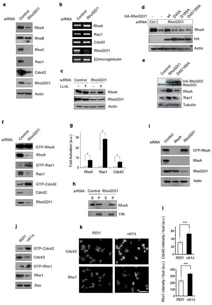
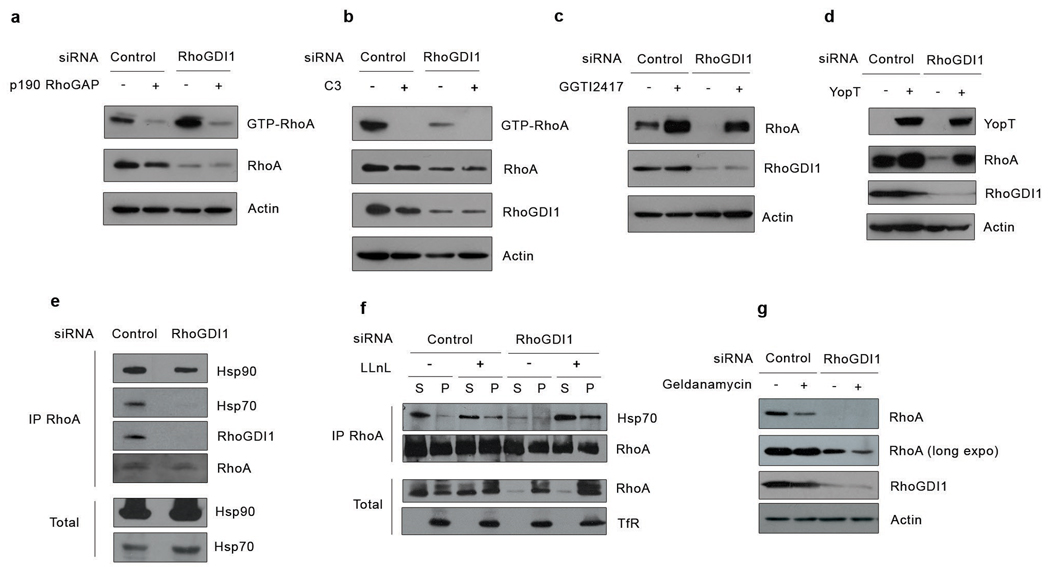
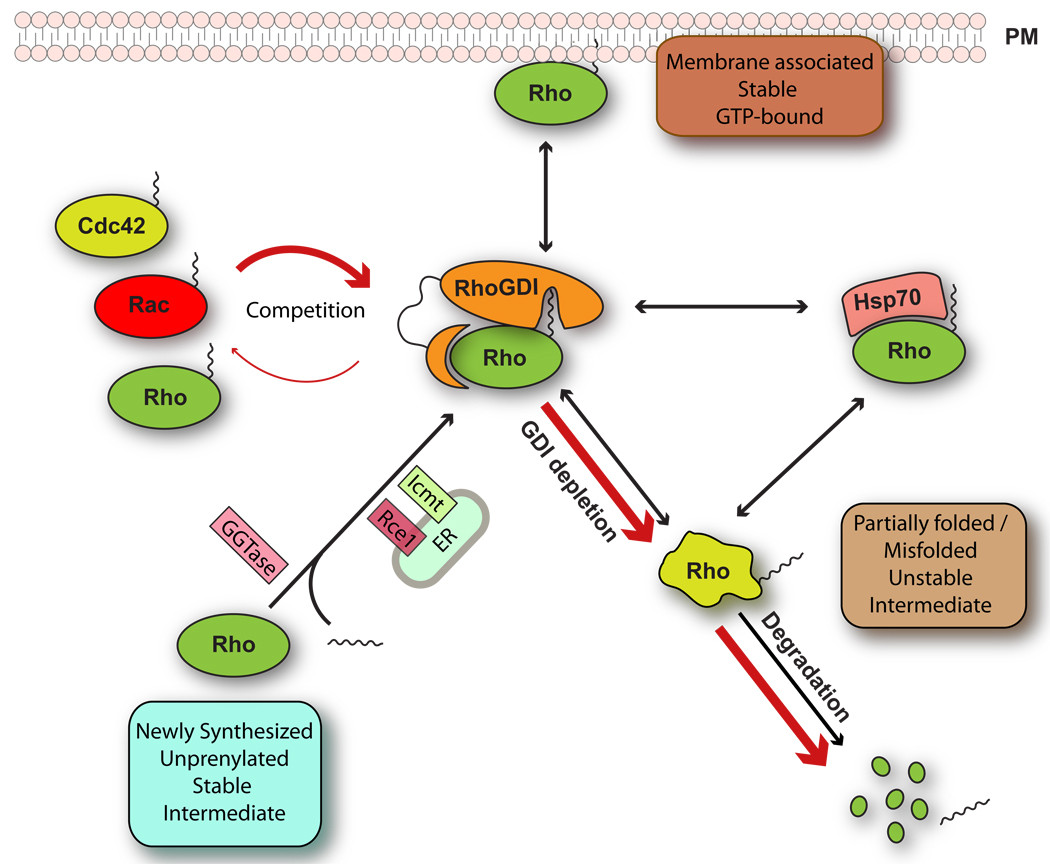
At steady state, most Rho GTPases are bound in the cytosol to Rho guanine nucleotide dissociation inhibitors (RhoGDIs). RhoGDIs have generally been considered to hold Rho proteins passively in an inactive state within the cytoplasm. Here we describe an evolutionarily conserved mechanism by which RhoGDI1 controls the homeostasis of Rho proteins in eukaryotic cells. We found that depletion of RhoGDI1 promotes misfolding and degradation of the cytosolic geranylgeranylated pool of Rho GTPases while activating the remaining membrane-bound fraction. Because RhoGDI1 levels are limiting, and Rho proteins compete for binding to RhoGDI1, overexpression of an exogenous Rho GTPase displaces endogenous Rho proteins bound to RhoGDI1, inducing their degradation and inactivation. These results raise important questions about the conclusions drawn from studies that manipulate Rho protein levels. In many cases the response observed may arise not simply from the overexpression itself but from additional effects on the levels and activity of other Rho GTPases as a result of competition for binding to RhoGDI1; this may require a re-evaluation of previously published studies that rely exclusively on these techniques.


Figures

Figure 1
Figure 1. RhoGDI1depletion triggers both degradation and activation of Rho proteins in eukaryotic cells
(a) Lysates from control or RhoGDI1 siRNA transfected HeLa cells were resolved by SDS-PAGE and analyzed by Western blotting. (b) Total RNAs were purified from control or RhoGDI1 siRNA transfected cells. RT-PCR was performed on DNAse I treated RNA using specific primers. RT-PCR products were resolved by agarose gel electrophoresis. (c) Lysates from control or RhoGDI1 siRNA transfected HeLa cells treated with LLnL for 8 hours were resolved by SDS-PAGE and analyzed by Western blotting. (d) HeLa cells were co-transfected with control or RhoGDI1 siRNA and HA-tagged RhoGDI1 RNAi resistant mutants. Cell lysates were resolved by SDS-PAGE and analyzed by Western blotting. All results are representative of at least 3 independent experiments. Note that the degradation of RhoA in RhoGDI1-depleted cells is rescued by expression of RhoGDI1 resistant to the siRNA, but not by RhoGDI1 mutants that do not bind Rho GTPases. (e) Lysates from HeLa cells transfected with HA-tagged wt RhoGDI1 or RhoGDI1 D45/185A were resolved by SDS-PAGE and analyzed by Western blotting. All siRNA experiments were analyzed at 72 h after transfection. For overexpression experiments, cells were transfected with the indicated cDNA 48 h after siRNA transfection and incubated for an additional 24 h. (f) HeLa cells were transfected with control or RhoGDI1 siRNA for 72 h. Active Rho GTPases were pulled-down from cell lysates with GST-RBD or GST-PBD beads. Bound proteins and total cell lysates were resolved by SDS-PAGE and analyzed by Western blotting. (g) Quantitation of the activation of RhoA, Rac1 and Cdc42 in RhoGDI1-depleted cells (black bars) relative to control cells (white bars). * p=0.0108, p=0.0146 and p=0.0229 respectively for RhoA, Rac1 and Cdc42 in a two-tail unpaired student’s t test with error bars representing standard error of the mean (s.e.m.) from three independent experiments. (h) Control or RhoGDI1 siRNA transfected cells were fractionated into cytosol and membrane fractions, resolved by SDS-PAGE and analyzed by Western blotting. TfR stands for transferrin receptor. (i) HeLa cells were infected with a RhoA miR shRNA-expressing adenovirus or transfected with RhoGDI1 siRNA for 72 h. Active RhoA was pulled-down with GST-RBD. Total cell lysates and GST-RBD bound proteins were resolved by SDS-PAGE and analyzed by Western blotting. (j) Active Rho GTPases from wild-type or Rdi1d yeast strains were pulled-down with GTP-RBD or GST-PBD beads. Bound proteins and total cell lysates were resolved by SDS-PAGE and analyzed by Western blotting. The Sso protein is used as loading control. (k) Immunofluorescence staining of Cdc42 and Rho1 in control wild-type (RDI1) and rdi1Δ strains. Scale bar is 8µm. (l) Quantitation of the intensity of polarized bud staining in control wild-type (RDI1) and rdi1Δ strains. Data were analyzed by two-tail student’s t test (p < 0.005) with error bars representing standard deviation from three experiments.
Figure 2
Figure 2. RhoGDI1 depletion impairs cell migration
(a) Time-course analysis of closure of a wound generated on a confluent monolayer of control or RhoGDI1 siRNA transfected HeLa cells. The graph depicts the wound area with error bars representing standard deviation (n=10). (b) Time-course analysis of closure of a wound generated on a confluent monolayer of control or RhoGDI1 siRNA transfected WM2664 melanoma cells. The graph depicts the wound area with error bars representing standard deviation (n=6). (c) Analysis of the velocity of control or RhoGDI1 siRNA transfected WM2664 melanoma cells. Data were analyzed by two-tailed unpaired student’s t test (p= 1.72×10⁻¹⁴) with error bars representing standard error of the mean (s.e.m.) from respectively n=28 and n=56 cells. The average speed of control and RhoGDI1 knock-down cells was 12.74 µm/min and 5.64 µm/min respectively. (d) WM2664 melanoma cells were transfected with control or RhoGDI1 siRNA for 72h. Active Rac1 and active RhoA were pulled-down from cell lysates with GST-PBD or GST-RBD beads respectively. Bound proteins and total cell lysates were resolved by SDS-PAGE and analyzed by Western blotting. (e) Cell lysates of control or RhoGDI1 siRNA transfected HeLa cells were resolved by SDS-PAGE and analyzed by Western blotting. Notice that Rho protein effectors are not activated despite the strong activation of RhoA and Rac. (f) Images extracted from supplementary information movie 1 depicting one WM2664 melanoma cell transfected with fluorescently labeled RhoGDI1 siRNA (red arrowhead) and two WM2664 cells transfected with unlabelled control siRNA (white and black arrowheads) migrating over time. Panel on right shows an image taken at t0 used to identify the cell transfected with the fluorescently labelled siRNA. Scale bar is 40 µm. (g) Representative XY migration tracks of control (left) or RhoGDI1 (right) siRNA transfected WM2664 melanoma cells. The positions of the cells were recorded every 5 minutes over a period of 2 hours (n=10). (h) HeLa cells transfected with control or RhoGDI1 siRNA for 72 h were fractionated into cytosolic or membrane fractions. Membrane fractions were further separated into plasma membrane (PM) and ER membrane (ER) by centrifugation on an iodixanol density gradient. Each fraction was analyzed by SDS-PAGE and Western blotting. (i) Densitometric analysis of the relative intensity of RhoA, Rac1 and Cdc42 bands in each fraction. Notice that upon RhoGDI1 silencing, the GTPases essentially disappear from the plasma membrane fractions.
Figure 3
Figure 3. Rho family GTPase degradation following RhoGDI1 depletion does not require activation of the Rho protein, but depends upon their geranylgeranylation and involves the molecular chaperone machinery
(a) HeLa cells were co-transfected with control or RhoGDI1 siRNA and p190RhoGAP cDNA. Active Rho GTPases were pulled-down from cell lysates with GST-RBD or GST-PBD beads. Bound proteins and total cell lysates were resolved by SDS-PAGE and analyzed by Western blotting. (b) HeLa cells were transfected with control or RhoGDI1 siRNA and treated with cell permeable C3 toxin. Active Rho GTPases were pulled-down from cell lysates with GST-RBD or GST-PBD beads. Bound proteins and total cell lysates were resolved by SDS-PAGE and analyzed by Western blotting. (c) HeLa cells were transfected with control or RhoGDI1 siRNA and treated with the geranylgeranyl transferase inhibitor GGTI 2417. Cell lysates were resolved by SDS-PAGE and analyzed by Western blotting. (d) HeLa cells were co-transfected with control or RhoGDI1 siRNA and a cDNA encoding the bacterial protease YopT. Cell lysates were resolved by SDS-PAGE and analyzed by Western blotting. (e) HeLa cells were transfected with control or RhoGDI siRNA and RhoA was immunoprecipitated from cell lysates. Immunoprecipitated proteins were resolved by SDS-PAGE and analyzed by Western blotting. (f) HeLa cells transfected with control or RhoGDI1 siRNA, with or without the proteasome inhibitor LLnL, were fractionated into cytosolic or membrane fractions and RhoA was immunoprecipitated from cytosolic and membrane fractions. To ensure that similar amounts of RhoA were immunoprecipitated, immunoprecipitations were performed with limiting amount of antibody and saturating amounts of cell lysate. Immunoprecipitated proteins and cell lysates were resolved by SDS-PAGE and analyzed by Western blotting. (g) HeLa cells were transfected with control or RhoGDI1 siRNA and treated with geldanamycin for 12 hours. Cell lysates were resolved by SDS-PAGE and analyzed by Western blotting. All results are representative of at least 3 independent experiments. A short and long exposure of the RhoA blot is shown. All siRNA experiments were analyzed at 72 h after transfection. For overexpression experiments, cells were transfected with the indicated cDNA 48 h after siRNA transfection and incubated for an additional 24 h.
Figure 4
Figure 4. Competitive interactions with RhoGDI1 regulate the levels and activities of Rho proteins
(a) HeLa cells were infected with a RhoA miR shRNA adenovirus for 72 h. Cell lysates were resolved by SDS-PAGE and analyzed by Western blotting. Note the increased levels of Rac1 and Cdc42 in cells from which RhoA has been depleted. (b) HeLa cells were transfected with myc-tagged Rho GTPases and endogenous RhoGDI1 was immunoprecipitated. Immunoprecipitated proteins and cell lysates were resolved by SDS-PAGE and analyzed by Western blotting. HeLa cells (c and d) or HEK 293 cells (e, f, g, h, i, j, k and l) were transfected with increasing amount of plasmid encoding for the indicated myc-tagged Rho GTPases. After 24 h, cell lysates were resolved by SDS-PAGE and analyzed by Western blotting. (m) HEK 293 cells were transfected with myc-tagged Cdc42 expressing plasmid with or without HA-tagged RhoGDI1 expressing plasmid for 24 h. Cell lysates were resolved by SDS-PAGE and analyzed by Western blotting. (n, o, and p) HEK 293 cells were transfected with 5µg of myc-tagged Rho GTPase expression plasmid as indicated. After 24 h, active RhoA or active Rac1 were pulled-down from cell lysates with GST-RBD or GST-PBD beads respectively. Bound proteins and total cell lysates were resolved by SDS-PAGE and analyzed by Western blotting. All results shown are representative of at least 3 independent experiments. Note that overexpression of one Rho family member decreases the expression and activity of the other Rho proteins.
Figure 5
Figure 5. RhoGDI regulates Rho protein homeostasis
Newly synthesized RhoGTPases are geranylgeranylated and posttranslationally modified in the ER. After geranylgeranylation, Rho proteins associate directly with RhoGDI which sequesters them as a soluble prenylated forming the cytosol and protects them from degradation. Upon depletion of RhoGDI1 or overexpression of Rho proteins, endogenous RhoGTPases are released to the cytosol where they exist as short-lived unstable intermediates that are partially folded or misfolded (red arrows). These can bind to the chaperone complex or be targeted for degradation if they are unable to fold properly. In the absence of GDI, newly synthesized Rho proteins cannot be delivered to the plasma membrane and accumulate in the ER. At steady state, this unstable intermediate is not detected and RhoGTPases are for the most part either associated with cell membranes or bound to GDI, with only a small fraction associated with the chaperone system. Abbreviations used: ER, endoplasmic reticulum; PM, plasma membrane; GGTase, geranylgeranyl transferase; Rce1, prenyl-protein specific protease; lcmt, isoprenylcysteine carboxyl methyltransferase.

References

    1. DerMardirossian C, Bokoch GM. GDIs: central regulatory molecules in Rho GTPase activation. Trends Cell Biol. 2005;15:356–363. - PubMed
    1. Dransart E, Morin A, Cherfils J, Olofsson B. Uncoupling of inhibitory and shuttling functions of rho GDP dissociation inhibitors. J Biol Chem. 2005;280:4674–4683. - PubMed
    1. Gorovoy M, et al. RhoGDI-1 modulation of the activity of monomeric RhoGTPase RhoA regulates endothelial barrier function in mouse lungs. Circ Res. 2007;101:50–58. - PubMed
    1. Abe M, Qadota H, Hirata A, Ohya Y. Lack of GTP-bound Rho1p in secretory vesicles of Saccharomyces cerevisiae. J Cell Biol. 2003;162:85–97. - PMC - PubMed
    1. Tong Z, et al. Adjacent positioning of cellular structures enabled by a Cdc42 GTPase-activating protein-mediated zone of inhibition. J Cell Biol. 2007;179:1375–1384. - PMC - PubMed

Publication types

MeSH terms

Substances