. 2010 Jun 17;29(24):3545-53.
doi: 10.1038/onc.2010.111. Epub 2010 Apr 19.

TWISTing stemness, inflammation and proliferation of epithelial ovarian cancer cells through MIR199A2/214

Affiliations

TWISTing stemness, inflammation and proliferation of epithelial ovarian cancer cells through MIR199A2/214

G Yin et al. Oncogene. .

Abstract

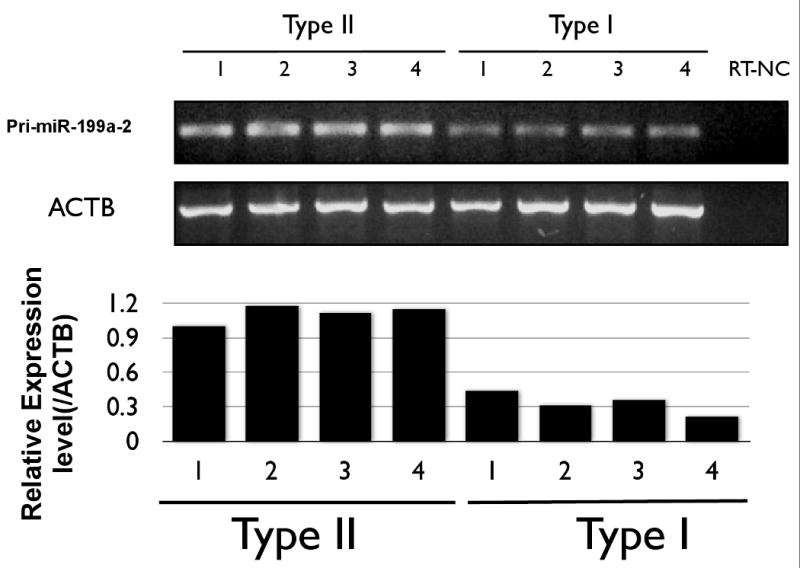
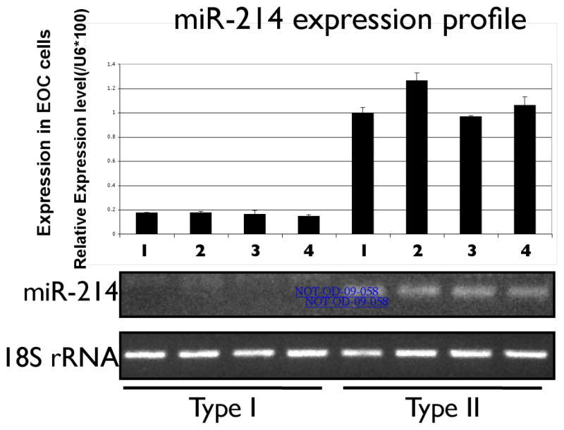
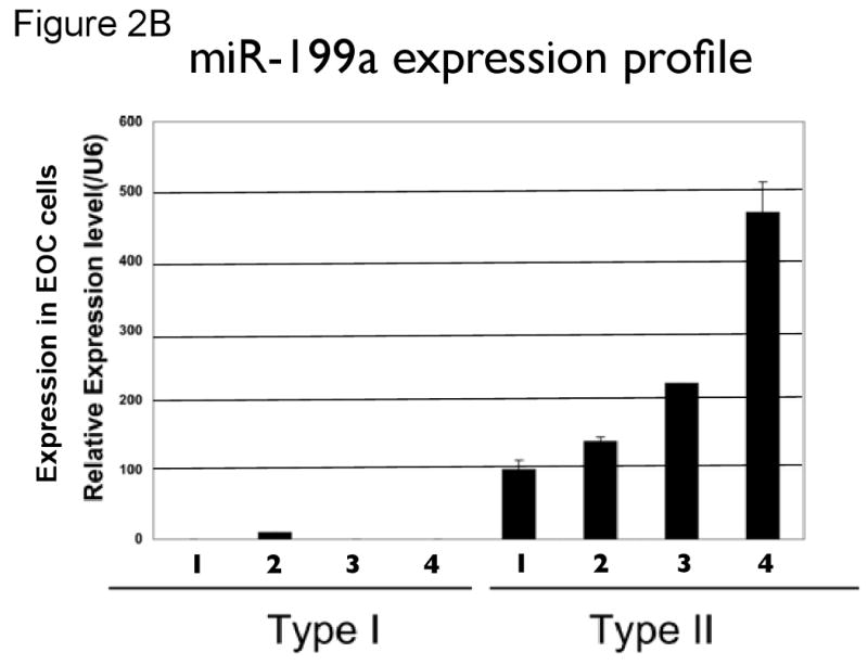
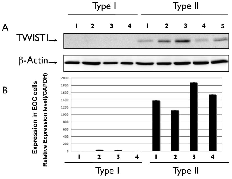
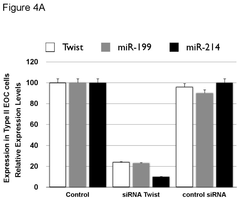
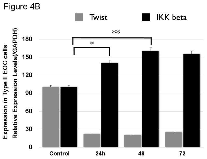
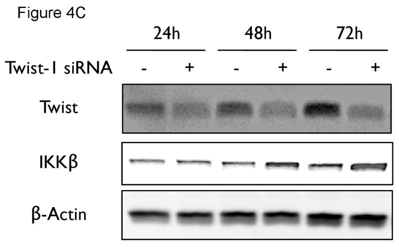
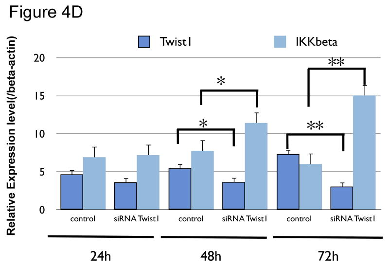
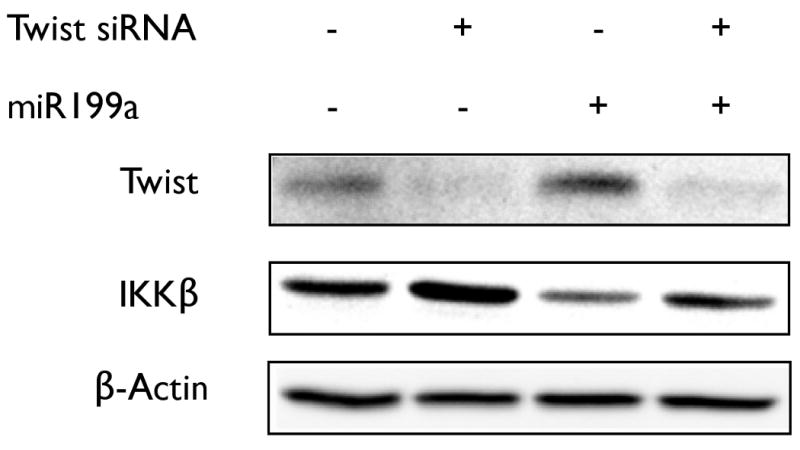
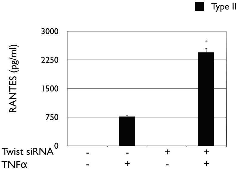
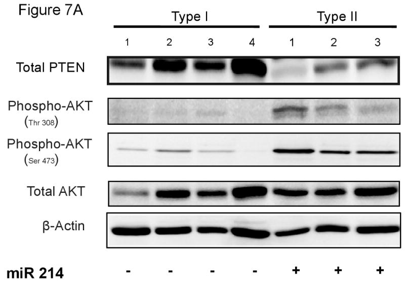
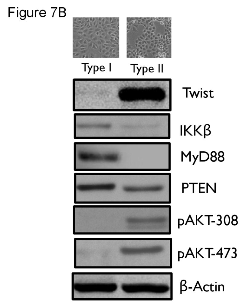
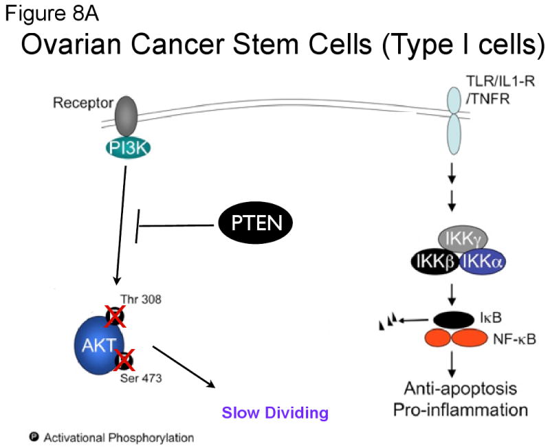
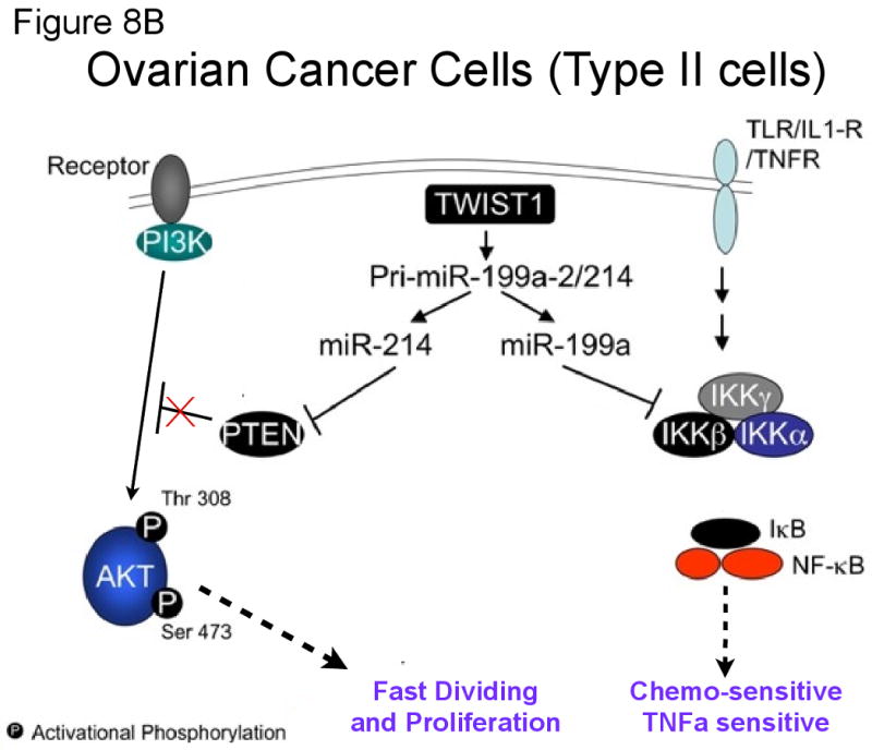
Cancer stem cells are responsible for sustaining the tumor and giving rise to proliferating and progressively differentiating cells. However, the molecular mechanisms regulating the process of cancer stem cell (CSC) differentiation is not clearly understood. Recently, we reported the isolation of the epithelial ovarian cancer (EOC) stem cells (type I/CD44+). In this study, we show that type I/CD44+ cells are characterized by low levels of both miR-199a and miR-214, whereas mature EOC cells (type II/CD44-) have higher levels of miR-199a and miR-214. Moreover, these two micro RNAs (miRNAs) are regulated as a cluster on pri-miR-199a2 within the human Dnm3os gene (GenBank FJ623959). This study identify Twist1 as a regulator of this unique miRNA cluster responsible for the regulation of the IKKbeta/NF-kappaB and PTEN/AKT pathways and its association of ovarian CSC differentiation. Our data suggest that Twist1 may be an important regulator of 'stemness' in EOC cells. The regulation of MIR199A2/214 expression may be used as a potential therapeutic approach in EOC patients.


Figures

Figure 1
Figure 1
Differential expression of pri-miR-199a-2 (from MIRN199A2) in EOC cells. Levels of pri-miR-199a-2 was evaluated in a panel of Type I/CD44+ and Type II/CD44- EOC cells using RT-PCR. ACTB: β-actin house keeping gene. RT-NC: Negative control for RT reaction.
Figure 2
Figure 2
Differential expression of miR-214 (a) and miR-199a (b) in EOC cells. Expression of the two miRNAs was determined in a panel of EOC cells by RT-PCR.
Figure 2
Figure 2
Differential expression of miR-214 (a) and miR-199a (b) in EOC cells. Expression of the two miRNAs was determined in a panel of EOC cells by RT-PCR.
Figure 3
Figure 3
Differential expression of Twist 1 in EOC cells. (a) Levels of Twist1 at the protein level was evaluated using Western blot analysis. (b) Twist1 mRNA levels were evaluated using RT-PCR.
Figure 4
Figure 4
Effect of Twist1 knockdown on levels of miR-199a, miR-214, and IKKβ. (a)Type II/CD44- EOC cells were transfected with Twist1 siRNA. Levels of Twist 1, miR-199a, and miR-214 were determined by RT-PCR. Note the significant decrease on miR-199a and miR-214 following Twist1 knockdown. No changes were observed in cells treated with control siRNA. (b) Type II/CD44- EOC cells were transfected with Twist1 siRNA and levels of IKKβ mRNA determined by RT-PCR in a time dependent manner. (c and d) Type II/CD44- EOC cells were transfected with Twist1 siRNA and levels of IKKβ determined by western blot analysis. Note that inhibition of Twist1 expression is associated with increase in IKKβ expression in a time dependent manner. *= p>0.05; **= p>0.001
Figure 4
Figure 4
Effect of Twist1 knockdown on levels of miR-199a, miR-214, and IKKβ. (a)Type II/CD44- EOC cells were transfected with Twist1 siRNA. Levels of Twist 1, miR-199a, and miR-214 were determined by RT-PCR. Note the significant decrease on miR-199a and miR-214 following Twist1 knockdown. No changes were observed in cells treated with control siRNA. (b) Type II/CD44- EOC cells were transfected with Twist1 siRNA and levels of IKKβ mRNA determined by RT-PCR in a time dependent manner. (c and d) Type II/CD44- EOC cells were transfected with Twist1 siRNA and levels of IKKβ determined by western blot analysis. Note that inhibition of Twist1 expression is associated with increase in IKKβ expression in a time dependent manner. *= p>0.05; **= p>0.001
Figure 5
Figure 5
Exogenous pre-miR-199a is able to reverse the effect of Twist1 knockdown on the levels of IKKβ. Type II/CD44- EOC cells were transfected with siRNA for Twist1. Afterwards, cells were incubated in the presence or absence of pre-miR-199a for additional 24 h and IKKβ expression was determined by Western blot analysis. Note that the presence of miR199a is able to reverse the effect of Twist1 inhibition.
Figure 6
Figure 6
Inhibition of Twist1 in Type II/CD44- EOC cells can reactivate the NF-κB pathway. Type II/CD44- EOC cells were transfected with siRNA for Twist1. Afterwards, cells were incubated in the presence or absence of TNFα (10 nM) for additional 24 hours. Levels of RANTES was determined in the supernatant. Note the significant increase in RANTES secretion in Type II/CD44- cells following Twist1 inhibition. Graph shown is representative of three independent experiments done in triplicate. * p=0.001
Figure 7
Figure 7
Expression of miR-214 correlates with the expression of PTEN and AKT phosphorylation status.(a) Levels of PTEN and two AKT phosphorylation sites were evaluated in a panel of Type I/CD44+ and Type II/CD44- EOC cells by western blot. Note the correlation between levels of PTEN, AKT phosphorylation and miR-214 expression. (b) Type I/CD44+ EOC cells were induced to differentiate in vitro into Type II/CD44- EOC cells and levels of Twist1, IKKβ, MyD88, PTEN, and pAKT was determined by western blot.
Figure 7
Figure 7
Expression of miR-214 correlates with the expression of PTEN and AKT phosphorylation status.(a) Levels of PTEN and two AKT phosphorylation sites were evaluated in a panel of Type I/CD44+ and Type II/CD44- EOC cells by western blot. Note the correlation between levels of PTEN, AKT phosphorylation and miR-214 expression. (b) Type I/CD44+ EOC cells were induced to differentiate in vitro into Type II/CD44- EOC cells and levels of Twist1, IKKβ, MyD88, PTEN, and pAKT was determined by western blot.
Figure 8
Figure 8
Proposed model for the role of Twist1 in the ovarian cancer stem cells. (a) In the ovarian cancer stem cells, undetectable levels of Twist1 results in undetectable expression of hsa-miR-199a and hsa-miR-214, the inhibitors of IKKβ and PTEN, respectively. This leads to high expression of both IKKβ and PTEN. As a result, the ovarian cancer stem cells have constitutive NF-kB activity, which creates a pro-inflammatory and anti-apoptotic environment, and an inactive Akt pathway, leading to slow proliferation. (b) In the mature ovarian cancer cells, high levels of Twist1 leads to the expression of hsa-miR-199a and hsa-miR-214, which downregulates IKKβ and PTEN, respectively. As a result, mature/differentiated ovarian cancer cells do not have a constitutive NF-kB pathway, but instead have a highly active Akt pathway. These cells do not have a pro-inflammatory environment, are chemosensitive, and proliferate faster.
Figure 8
Figure 8
Proposed model for the role of Twist1 in the ovarian cancer stem cells. (a) In the ovarian cancer stem cells, undetectable levels of Twist1 results in undetectable expression of hsa-miR-199a and hsa-miR-214, the inhibitors of IKKβ and PTEN, respectively. This leads to high expression of both IKKβ and PTEN. As a result, the ovarian cancer stem cells have constitutive NF-kB activity, which creates a pro-inflammatory and anti-apoptotic environment, and an inactive Akt pathway, leading to slow proliferation. (b) In the mature ovarian cancer cells, high levels of Twist1 leads to the expression of hsa-miR-199a and hsa-miR-214, which downregulates IKKβ and PTEN, respectively. As a result, mature/differentiated ovarian cancer cells do not have a constitutive NF-kB pathway, but instead have a highly active Akt pathway. These cells do not have a pro-inflammatory environment, are chemosensitive, and proliferate faster.

Similar articles

Cited by

References

    1. Ahmed N, Thompson EW, Quinn MA. Epithelial-mesenchymal interconversions in normal ovarian surface epithelium and ovarian carcinomas: an exception to the norm. J Cell Physiol. 2007;213:581–588. - PubMed
    1. Alison MR, Murphy G, Leedham S. Stem cells and cancer: a deadly mix. Cell Tissue Res. 2008;331:109–124. - PubMed
    1. Alvarez-Garcia I, Miska EA. MicroRNA functions in animal development and human disease. Development. 2005;132:4653–4662. - PubMed
    1. Alvero AB, Chen R, Fu HH, Montagna M, Schwartz PE, Rutherford T, et al. Molecular phenotyping of human ovarian cancer stem cells unravels the mechanisms for repair and chemoresistance. Cell Cycle. 2009a;8:158–166. - PMC - PubMed
    1. Alvero AB, Fu HH, Holmberg J, Visintin I, Mor L, Marquina CC, et al. Stem-like Ovarian Cancer Cells can Serve as Tumor Vascular Progenitors. Stem Cells 2009b - PMC - PubMed

Publication types

MeSH terms

Associated data