. 2010 Mar 1;5(2):227-246.
doi: 10.2217/fnl.10.1.

Microglia in ischemic brain injury

Affiliations

Microglia in ischemic brain injury

Jonathan R Weinstein et al. Future Neurol. .

Abstract

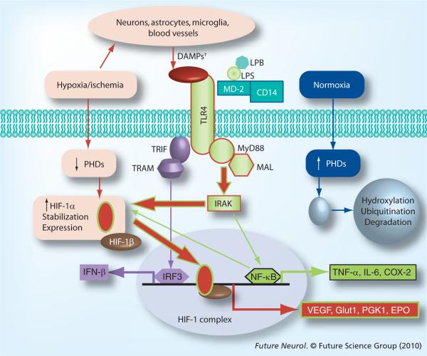
Microglia are resident CNS immune cells that are active sensors in healthy brain and versatile effectors under pathological conditions. Cerebral ischemia induces a robust neuroinflammatory response that includes marked changes in the gene-expression profile and phenotype of a variety of endogenous CNS cell types (astrocytes, neurons and microglia), as well as an influx of leukocytic cells (neutrophils, macrophages and T-cells) from the periphery. Many molecules and conditions can trigger a transformation of surveying microglia to microglia of an alerted or reactive state. Here we review recent developments in the literature that relate to microglial activation in the experimental setting of in vitro and in vivo ischemia. We also present new data from our own laboratory demonstrating the direct effects of in vitro ischemic conditions on the microglial phenotype and genomic profile. In particular, we focus on the role of specific molecular signaling systems, such as hypoxia inducible factor-1 and Toll-like receptor-4, in regulating the microglial response in this setting. We then review histological and novel radiological data that confirm a key role for microglial activation in the setting of ischemic stroke in humans. We also discuss recent progress in the pharmacologic and molecular targeting of microglia in acute ischemic stroke. Finally, we explore how recent studies on ischemic preconditioning have increased interest in pre-emptively targeting microglial activation in order to reduce stroke severity.


Figures

Figure 1
Figure 1. Induction of hypoxia-inducible factor-1 and its downstream gene targets in primary microglia
For determination of total cellular HIF1α. (A), wild-type (WT) primary mouse microglia (pMG) were serum-starved and then either unstimulated, stimulated with cobalt chloride (200 μM) or treated under hypoxic conditions (pO2 = 1%) for 6 h. Lysates were prepared and total cellular HIF-1α ELISA carried out as per manufacturers' instructions (R&D Systems, MN, USA). In (B & C), serum-starved pMG were exposed to either normoxic (pO2 = 21%)/normoglycemic (5 mM glucose) conditions or hypoxic (pO2 = 1%)/hypoglycemic (250 μM glucose) conditions ± 100 EU/ml LPS (Associates of Cape Cod, Inc., MA, USA) for 24 h. Total RNA was extracted and samples were processed by qRT-PCR for steady-state levels of VEGF and Glut-1 mRNA as described [42]. In (A−C), each bar in the graph shows the result of a representative individual experiment (n = 2 for all conditions). HIF: Hypoxia inducible factor; LPS: Lipopolysaccharide.
Figure 2
Figure 2. Microarray analyses on wild type and TLR4−/− primary mouse microglia following exposure to experimental or control conditions
WT and TLR4−/− primary mouse microglia (pMG) were exposed to hypoxic/hypoglycemic (`ischemic') or normoxic/normoglycemic (`control') conditions for 24 h in triplicate. All cells were then exposed to 24 h of normoxic/normoglycemic (`reperfusion') conditions. RNA was extracted, analyzed and processed for cDNA synthesis and cDNAs were hybridized to a Mouse Gene 1.0 ST Array and raw data processed, normalized and analyzed as described [43,44]. Individual genes with evidence for significant differential expression between strains (WT vs TLR4−/−) and/or conditions (ischemia–reperfusion vs control) were identified as described [45]. Comparisons showing distinct and overlapping subsets of regulated individual genes are presented as Venn diagrams (A). Individual genes were sorted into biological processes (BP) using gene-set enrichment analyses (GSEA), as described [46,47]. Differential ischemia-induced regulation of selected BP categories between strains is presented here as a bar graph (B). All microarray data have been deposited in the Gene Expression Omnibus Database under accession number GSE18602. KO: Knockout; TLR: Toll-like receptor; WT: Wild-type.
Figure 3
Figure 3. Select TLR4 and HIF-1α signaling pathways in microglia
Pathways associated with normoxia, hypoxia/ischemia and TLR4 activation by LPS are outlined in blue, red or green, respectively. DAMPS include HSP, HMGB1 and other endogenous ligands for TLR4. DAMP: Danger-associated molecular pattern; HIF: Hypoxia inducible factor; LPB: LPS-binding protein; LPS: Lipopolysaccharide; PHD: Prolylhydroxylase protein; TLR: Toll-like receptor; TRAM: TRIF-related adaptor molecule; TRIF: Toll–IL-1 receptor domain-containing adaptor factor.
Figure 4
Figure 4. Summary of findings and model of the ischemic penumbra
(A) Representative confocal micrographs of the healthy rat cortex, showing the dense network of ramified microglia whose processes are in close apposition to the neurons. Scale bars: left, 200 μm; right, 10 μm. A higher-magnification image shows a typical microglial cell with processes abutting the neuronal cell body (near top) and wrapping around the axonal processes. Microglia are labeled with a rabbit polyclonal antibody against a membrane protein, IBA1, and an Alexa 488-conjugated goat anti-rabbit secondary antibody (green). Neurons are labeled with a mouse monoclonal MAP2 antibody and Cy3-conjugated goat antimouse secondary antibody (red). (B) After a stroke, an inflammatory response develops over time, microglia become activated (their processes retract and they migrate to the damage site), and neuronal damage propagates into the surrounding tissue, the ischemic `penumbra'. Note that the colors are reversed from (A); microglia (red) are labeled with IBA1 antibody and a Cy3-conjugated secondary antibody, whereas rat cortical neurons (green) are labeled with microtubule-associated protein-2 and a FITC-conjugated secondary antibody. Author's interpretation [49]: “In these representative confocal micrographs from the rat brain after a stroke, microglia (green) are ramified and densely distributed in the uninjured contralateral hemisphere (top image), but retract their processes and eventually become rounded up as they progressively activate on the damaged ipsilateral side (bottom two images). In this study, using an in vitro model of the stroke penumbra, we identified several events: glutamate, released by the oxygen–glucose deprivation-stressed neuron–astrocyte cultures, reached sufficient concentrations to stimulate microglial mGluRIIs; this stimulation was blocked by the selective mGluRII antagonists, EGLU or LY341495; microglial activation was accompanied by activation of NF-κB, not p38 MAPK; activated microglia produced and released TNF-α; TNF-α interacted with the target neurons, activating their p55/TNF1 receptors, as judged by activation of caspase-8. Neurotoxicity was reduced by scavenging TNF-α with sTNFR1; the target neurons died by apoptosis, as judged by caspase-3 activation and TUNEL; caspase 3 activation was required for excess neuron killing, because it was prevented by the caspase-3 inhibitor, DEVD-CHO, and by the broad-spectrum caspase inhibitor, Boc-D-FMK.” mGluR: Metabatropic glutamate receptor II; sTNFR1: Soluble TNF-α receptor 1; TNFR: TNF receptor. Adapted with permission from [49].
Figure 5
Figure 5. Temporal course of infiltration of inflammatory cells in experimental models of stroke
Activated microglia appear in the ipsilateral hemisphere early after focal ischemia, followed by an influx of neutrophils into the parenchyma. Macrophage infiltration after ischemia is a later event in most experimental paradigms.
Figure 6
Figure 6. 11C-(R)-PK11195 radiolabeled activated microglia map to the peri-infarct zone in the sub-acute phase of stroke in humans
Case shown depicts PET and MRI imaging findings at 2, 13 and 30 days postischemic infarction in the left basal ganglia of a 43-year-old woman. A color binding potential scale referring to the left hand column is provided. The right hand column demonstrates significant binding potential within the ischemic core (purple) and peri-infarct penumbra (green). Adapted with permission from [110].

References

    1. Mohr JP, Choi DW, Grotta JC, Weir B, Wolf PA. Stroke: Pathophysiology, diagnosis, and management. Fourth Edition Churchill Livingstone (an Imprint of Elsevier, Inc.); 2004.
    1. Dirnagl U, Iadecola C, Moskowitz MA. Pathobiology of ischaemic stroke: an integrated view. Trends Neurosci. 1999;22(9):391–397. - PubMed
    1. Green RA, Odergren T, Ashwood T. Animal models of stroke: do they have value for discovering neuroprotective agents? Trends Pharmacol. Sci. 2003;24(8):402–408. - PubMed
    1. Stroke Therapy Academic Industry Roundtable (STAIR) Preclinical Recommendations. Writing Committee composed of the following members: Finklestein SP, Fisher M (Chairman), Furlan AJ, Goldstein LB, Gorelick PB, Kaste M, Lees KR, Traystman RJ Recommendations for standards regarding preclinical neuroprotective and restorative drug development. Stroke. 1999;30(12):2752–2758.

    1. Becker KJ. Targeting the central nervous system inflammatory response in ischemic stroke. Curr. Opin. Neurol. 2001;14(3):349–353. - PubMed