. 2008 Mar 3:1:9-15.
doi: 10.4137/vrt.s563.

Identification of Rotavirus VP6-Specific CD4+ T Cell Epitopes in a G1P[8] Human Rotavirus-Infected Rhesus Macaque

Affiliations

Identification of Rotavirus VP6-Specific CD4+ T Cell Epitopes in a G1P[8] Human Rotavirus-Infected Rhesus Macaque

Wei Zhao et al. Virology (Auckl). .

Abstract

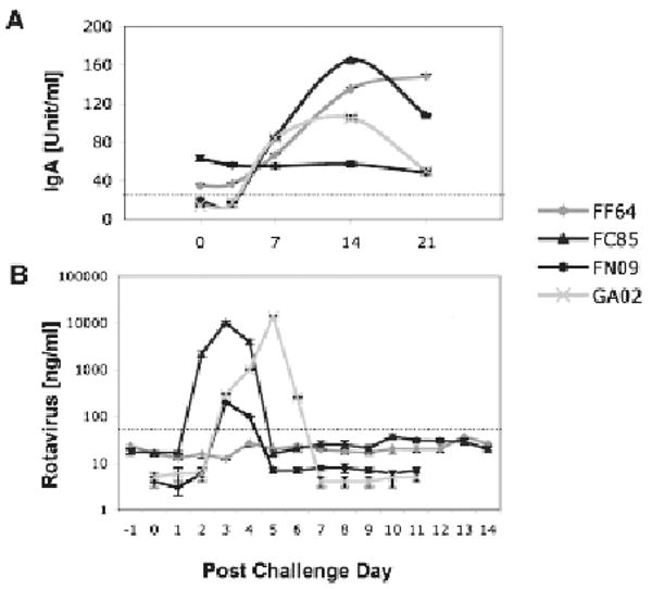
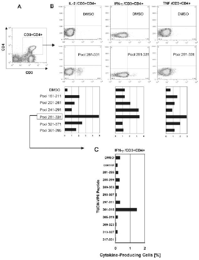
A non-human primate model was used to evaluate its potential for identification of rotavirus viral protein 6 (VP6) CD4+ T cell epitopes. Four juvenile rhesus macaques were inoculated with a mixed inoculum (G1P[8] and G9P[8]) of human rotaviruses. Infection accompanied by G1P[8] shedding was achieved in the two macaques that had no rotavirus immunoglobulin A (IgA) in plasma. To measure the interferon gamma (IFN-γ) and tumor necrosis factor (TNF) anti-viral cytokines produced by peripheral CD4+ cells that recognize VP6 epitopes, whole blood cells from one infected macaque were stimulated in vitro with VP6 peptides. Stimulation with peptide pools derived from the simian rotavirus VP6(161-395) region revealed reactivity of CD4+ T cells with the VP6(281-331) domain. A VP6(301-315) region was identified as the epitope responsible for IFN-γ production while a broader VP6(293-327) domain was linked to TNF production. These results suggest that human rotavirus-infected macaques can be used for identification of additional epitopes and domains to address specific questions related to the development of pediatric vaccines.


Figures

Figure 1
Figure 1
Rotavirus plasma IgA (A) and fecal shedding (B) are shown upon experimental challenge of 4 juvenile macaques with human rotavirus. Dotted lines represent cut-offs between negative and positive measurements—as previously established by average values corresponding to populations of control (negative and positive) macaques. Immunoassays used to measure the rotavirus plasma IgA and fecal shedding were described in detail elsewhere (Sestak et al. 2004; McNeal et al. 2005; Zhao et al. 2005).
Figure 2
Figure 2
Rotavirus VP6 epitope mapping was performed with peripheral blood cells collected at peak of virus infection (PCD 7) from GA02 macaque. Stimulation was performed in vitro with overlapping VP6 15-mer peptide pools corresponding to the TUCH rotavirus (Maecker et al. 2001; Jaimes et al. 2005). Following staining with appropriate FACS antibodies, analysis of intracellular cytokine production was performed (Sestak et al. 2004; Pahar et al. 2006). Gating was done through populations of CD3+CD4+ T cells (A). The amino acid sequence ranges are shown for each pool (B). Highest production of IL-2, TNF and IFN-γ cells was found upon stimulation with the VP6281–331 pool although some cytokine production was also seen upon stimulation with other VP6 pools, primarily for IFN-γ after stimulation with the VP6241–291 peptide pool. DMSO values indicate baseline levels. Individual peptides from the VP6281–331 pool were used to stimulate peripheral blood cells collected at PCD 14 for their ability to induce production of IFN-γ (C). The highest IFN-γ production following stimulation with VP6301–315 peptide is shown (C).

References

    1. Banos DM, Lopez S, Arias CF, Esquivel FR. Identification of a T-helper cell epitope on the rotavirus VP6 protein. J Virol. 1997;71:419–26. - PMC - PubMed
    1. Choi AH, Basu M, McNeal MM, Flint J, VanCott JL, Clements JD, Ward RL. Functional mapping of protective domains and epitopes in the rotavirus VP6 protein. J Virol. 2000;74:11574–80. - PMC - PubMed
    1. Conner ME, Gilger MA, Estes MK, Graham DY. Serologic and mucosal immune response to rotavirus infection in the rabbit model. J Virol. 1991;65:2562–71. - PMC - PubMed
    1. Das BK, Gentsch JR, Cicirello HG, Woods PA, Gupta A, Ramachandran M, Kumar R, Bahn MK, Glass RI. Characterization of rotavirus strains from newborns in New Delhi, India. J Clin Microbiol. 1994;32:1820–2. - PMC - PubMed
    1. Doxiadis GGM, Otting N, de Groot NG, Bontrop RE. Differential evolutionary MHC class II strategied in humans and rhesus macaques: relevance for biomedical studies. Immunol Rev. 2001;183:76–85. - PubMed