Review
. 2010 Apr 26;49(19):3280-94.
doi: 10.1002/anie.200904359.

Gold nanoparticles for biology and medicine

Affiliations
Review

Gold nanoparticles for biology and medicine

David A Giljohann et al. Angew Chem Int Ed Engl. .

Abstract

Gold colloids have fascinated scientists for over a century and are now heavily utilized in chemistry, biology, engineering, and medicine. Today these materials can be synthesized reproducibly, modified with seemingly limitless chemical functional groups, and, in certain cases, characterized with atomic-level precision. This Review highlights recent advances in the synthesis, bioconjugation, and cellular uses of gold nanoconjugates. There are now many examples of highly sensitive and selective assays based upon gold nanoconjugates. In recent years, focus has turned to therapeutic possibilities for such materials. Structures which behave as gene-regulating agents, drug carriers, imaging agents, and photoresponsive therapeutics have been developed and studied in the context of cells and many debilitating diseases. These structures are not simply chosen as alternatives to molecule-based systems, but rather for their new physical and chemical properties, which confer substantive advantages in cellular and medical applications.


Figures

Figure 1
Figure 1
Transmission electron microscopy imaging and measurements of gold nanoparticles in cells. A) Graph of number of gold nanoparticles per vesicle diameter for various nanoparticle sizes. B–F) TEM images of gold nanoparticles with sizes of 14, 30, 50, 74, and 100 nm, respectively, trapped inside vesicles of a HeLa cell. Adapted from Ref. [19], with permission from the American Chemical Society; Copyright 2006.
Figure 2
Figure 2
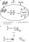
A) Schematic illustration of the release of DNA from a photocleavable AuNP complex (NP-PC) upon UV irradiation within the cell. B) Schematic presentation of light-induced surface transformation of NP-PC. Adapted from Ref. [35].
Figure 3
Figure 3
The synthesis of the oligonucleotide gold nanoconjugates: Alkanethiol-terminated oligonucleotides are added to citrate-stabilized AuNPs, thereby displacing the capping citrate ligands through formation of a gold–thiol bond. Subsequent addition of a salt shields repulsion between the strands, thus leading to a dense monolayer of oligonucleotides.
Figure 4
Figure 4
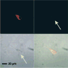
Fluorescent microscopy images of C166-EGFP cells incubated for 48 h with gold nanoconjugates functionalized with dual-fluorophore-labeled oligonucleotides (3′-Cy3 and 5′-Cy5.5) only reveal fluorescence from Cy5.5 (706–717 nm, upper left). Negligible fluorescence is observed in the emission range of Cy3 (565–615 nm, upper right). Transmission and composite overlay images are shown in the lower left and lower right quadrants, respectively. The arrows indicate the location of the cell. Adapted from Ref. [25], with permission from the American Association for the Advancement of Science; Copyright 2006.
Figure 5
Figure 5
A) Representative Western blots showing the expression of glyceradlehyde 3-phosphate dehydrogenase (GAPDH) in HeLa cells treated with various concentrations and compositions of the gold nanoconjugates. GAPDH expression is reduced in a dose- and sequence-dependent manner. α-Tubulin is shown as the loading control. B) Relative decrease in GAPDH expression in HeLa cells. α-Tubulin was used as a loading control and for subsequent normalization of GAPDH knockdown. The error bars represent the standard deviation from at least three Western blots. Adapted from Ref. [102], with permission from the National Academy of Sciences; Copyright 2008.
Figure 6
Figure 6
“Nanoflares” are gold nanoconjugates functionalized with oligonucleotide sequences complementary to a specific nucleic acid target (messenger RNA) hybridized to short fluorescent sequences. In the absence of a target the nanoflares are dark, because of quenching by the gold nanoparticle. In the presence of a target binding displaces the short flare through the formation of a longer (more energetically favorable) duplex. The result is a fluorescence signal inside the cell, which indicates the target has been detected. Scale bar: 20 μm. Adapted from Ref. [87], with permission from the American Chemical Society; Copyright 2007.
Figure 7
Figure 7
Images of nanoparticle–peptide complexes incubated with HepG2 cells for 2 h. Complexes were: A) nuclear localization peptide, B) receptor-mediated endocytosis peptide, C) adenoviral fiber protein, and D) both nuclear localization and receptor-mediated endocytosis peptides. Adapted from Ref. [23], with permission from the American Chemical Society; Copyright 2003.
Figure 8
Figure 8
Templated synthesis of spherical HDL nanoparticles through use of thiol-terminated peptides and the protein (APOA1). Adapted from Ref. [111], with permission from the American Chemical Society; Copyright 2009.

References

    1. Hayat MA. Colloidal gold: principles, methods, and applications. Academic Press; San Diego: 1989.
    1. Edwards PP, Thomas JM. Angew Chem. 2007;119:5576. - PubMed
    2. Angew Chem Int Ed. 2007;46:5480. - PubMed
    1. Daniel MC, Astruc D. Chem Rev. 2004;104:293. - PubMed
    1. Mirkin CA, Letsinger RL, Mucic RC, Storhoff JJ. Nature. 1996;382:607. - PubMed
    1. Park SY, Lytton-Jean AKR, Lee B, Weigand S, Schatz GC, Mirkin CA. Nature. 2008;451:553. - PubMed

Publication types

LinkOut - more resources