Review
. 2010 Aug 11;110(8):4820-38.
doi: 10.1021/cr900377t.

Polymorphism in Alzheimer Abeta amyloid organization reflects conformational selection in a rugged energy landscape

Affiliations
Free PMC article
Review

Polymorphism in Alzheimer Abeta amyloid organization reflects conformational selection in a rugged energy landscape

Yifat Miller et al. Chem Rev. .
Free PMC article
No abstract available


Figures

Figure 1
Figure 1
Polymorphism of full-length Aβ peptides observed in experiments. (A) Transmission electron microscopy (TEM) images of amyloid fibrils formed by the Aβ1−40 peptide, prepared by incubation of Aβ1−40 solutions under quiescent dialysis conditions. Reprinted with permission from ref (72). Copyright 2005 Science/AAAS. (B) Cryo electron microscopy (cryo-EM) of the structural persistence and morphological diversity of Aβ1−40 fibrils grown either in sodium borate (pH 7.8) at 22 °C or in phosphate-buffered saline (PBS) (pH 7.4) at 37 °C. Both samples encompass evidently more than one fibril morphology; Reprinted with permission from ref (73). Copyright 2009 Elsevier. (C) Electron microscopy (EM) images of morphologies of Aβ1−42 fibrils. Reprinted with permission from ref (15). Copyright 2005 National Academic of Sciences, USA.
Figure 2
Figure 2
Polymorphism of Aβ peptide fragments as observed in EM images. (A) Aβ1−28 fibrils, negatively stained, are periodically stained along some of their edges (indicated by arrows). Fibril ends appear to splay or unfurl. Reprinted with permission from ref (75). Copyright 1987, directly permission from Dennis J. Selkoe. (B) Aβ10−23 filaments prepared in vitro. The filaments are irregularly twisted around each other. Reprinted with permission from ref (79). Copyright 1991 Elsevier. (C) Aβ26−33 and Aβ34−42 fibrils. Aβ26−33 fibrils are thin and uniform as compared to those of Aβ34−42 fibrils and appears to be comprised of fibril pairs. At low magnification, Aβ34−42 shows two types of assemblies: twisted fibrils and untwisted narrow fibrils. At higher magnification, twisted fibrils and periodic deposition of enhanced staining (indicated by arrows) are observed. Reprinted with permission from ref (101). Copyright 1990 American Chemical Society. (D) Aβ17−40 form protofibrils (top) and a mature fibril of Aβ17−42 (bottom). Reprinted with permission from ref (103). Copyright 2003 American Society for Biochemistry and Molecular Biology.
Figure 3
Figure 3
(A) EM images and X-ray diffraction patterns of cross-β ribbons for Aβ11−25 (I) and (III) and Aβ1−40 (II) and (IV). The measured widths of the fibrils are ∼5 nm for Aβ11−25 and ∼7 nm for Aβ1−40 (see arrows in I and II). These are wide-angle X-ray patterns; a* is the vertical fibril axis;. Reprinted with permission from ref (77). Copyright 2003 Elsevier. (B) Model of Aβ11−25: (I) Antiparallel organization. The 0.47 nm spacing (a-direction) is controlled by hydrogen bonding, and the molecules are ∼5 nm in length. (II) Direct stacking that would generate an orthorhombic unit cell. (III) Stacking with a slip of 0.697 nm (one β-sheet crimp; one structural repeat in the c-direction) parallel to the c axis. This stacking arrangement gives rise to monoclinic unit cell. Reprinted with permission from ref (77). Copyright 2003 Elsevier. (C) Illustration of cross-β arrangement in amyloids; the peptide backbone β-strands are perpendicular to the fibril axis (arrow).
Figure 4
Figure 4
Potential architectures of strand organizations in amyloid fibrils. Interactions (in dotted lines) are shown between the first layer (blue) and the second layer (red) for (A) parallel to parallel, face-to-face orientation; (B) parallel to parallel, face-to-back orientation; (C) parallel to antiparallel, face-to-face orientation; (D) parallel to antiparallel, face-to-back orientation; (E) antiparallel to antiparallel, face-to-face orientation, parallel to parallel between layers; (F) antiparallel to antiparallel, face-to-back orientation, parallel to parallel between layers; (G) antiparallel to antiparallel, face-to-back orientation, parallel to parallel between layers; and (H) antiparallel to antiparallel, face-to-back orientation, antiparallel to antiparallel between layers.
Figure 5
Figure 5
Illustration of Aβ25−35 in parallel (A) and antiparallel (B) arrangements. The sticks are peptide backbone, and the balls are Ile residues. Parts (C) and (D) illustrate the U-turn structures of Ma−Nussinov−Tycko (ref (46)) and Lührs et al. (ref (15)), respectively.
Figure 6
Figure 6
Polymorphism of the Aβ1−40 peptide based on different associations of the protofibrils. (A) A structural model of protofilament core topology of fibrils 1, 5, and 11, observed from Meinhardt et al.(73) Side view of the fibrils with two protofilament cores modeled into the density (top) and contoured density cross sections of the fibrils superimposed with two protofilament cores (bottom). Each protofilament core comprises a pair of two β-sheet regions: interface (yellow) and outside (blue). Reprinted with permission from ref (73). Copyright 2009 Elsevier. (B) Experiment-based structural models of Aβ9−40. (I) A ribbon presentation of the lowest-energy model for fibrils with twisted morphology. (II) Atomic representation, viewed down the fibril axis. Hydrophobic, polar, negatively charged, and positively charged amino acid side-chains are green, magenta, red, and blue, respectively. Backbone nitrogen and carbonyl oxygen atoms are cyan and pink. (III) Comparison of twisted (upper) and striated ribbon (lower) fibril morphologies in negatively TEM images. (IV) Atomic representation of a model for striated ribbon fibrils developed previously by Petkova et al., Reprinted with permission from ref (138). Copyright 2008 National Academic of Sciences, USA.
Figure 7
Figure 7
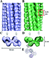
Variability in β2-microglobulin fibril structure. Reprinted with permission from ref (159). Copyright 2009 Elsevier. Three-dimensional reconstructions of the type A and type B forms of β2-microglobulin fibrils. Side views of an A-type fibril (A) and a B-type fibril (B). One dimeric density unit is indicated by a red box in (B). The directions of the half-fibrils are indicated by arrows below the maps. Cross sections of the A type (C) and B-type fibrils (D) show that the structures are formed of crescent-shaped units stacked back-to-back. (E) Superposed contour plots of the A (lilac) and B (green) repeat units, showing that the two fibril types have the same underlying organization that differs only in the orientation of the two stacks, either parallel or antiparallel.
Figure 8
Figure 8
Illustration of the organization of the hydrophobic residues (black) and the charged residues (red), and their charge interactions (brown) in parallel and antiparallel alignment for (A) Aβ10−35, (B) Aβ16−22, and (C) Aβ34−42 fragments.
Figure 9
Figure 9
Various conformations of Zn2+−Aβ monomer complexes: (A) NMR structural model for Zn2+−Aβ1−16 (ref (252)), (B) NMR structural model for human Zn2+−Aβ1−28 (ref (257)), (C) NMR structural model for rat Zn2+−Aβ1−28 (ref (257)), and (D) X-ray spectroscopy model of Zn2+ binding to His13 and His14 of 2 adjacent Aβ peptides (ref (258)).
Figure 10
Figure 10
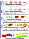
Polymerization of Aβ peptides involves continuous hierarchical redistributions of the polymorphic ensembles on a rugged energy landscape. For illustration, we present five aggregation phases starting from the native states and ending in the fibril forms. The arrow on the left indicates the evolution of the aggregated states with time: (A) The conformational ensemble of the “normal” folded monomers (three schematic figures in the middle) and partially disordered or unfolded states (the right and the left schematic figures). (B) Under certain conditions, folded monomers assemble toward polymorphic seeds or partially aggregated states (green, blue, and yellow boxes), redistributing the conformational ensemble. The native disordered or unfolded states (red schematic figures) may still have a large population. (C) “Critical intermediates” may be different partially aggregate states (green, blue, and yellow boxes) and very early stage of small oligomers (two right schematic figures). (D) Certain “critical intermediates” can lead to both different amyloid seeds (three left schematic figures) and different soluble ADDL-like oligomers (orange balls). The soluble ADDL-like oligomers are prior to amyloidogenic protofibril formation. Polymorphic seeds and polymorphic soluble oligomers present different toxic mechanisms (discussed in section 7). (E) The different fibril forms are the most stable states of the Aβ peptide amyloid: protofilament fragments (blue), protofibril forms (yellow and green), and mature fibril (left schematic figure).

Similar articles

Cited by

References

    1. Fowler D. M.; Koulov A. V.; Balch W. E.; Kelly J. W. Trends Biochem. Sci. 2007, 32, 217. - PubMed
    1. Maury C. P. J. Intern. Med. 2009, 265, 329. - PubMed
    1. Maji S. K.; Perrin M. H.; Sawaya M. R.; Jessberger S.; Vadodaria K.; Rissman R. A.; Singru P. S.; Nilsson K. P.; Simon R.; Schubert D.; Eisenberg D.; Rivier J.; Sawchenko P.; Vale W.; Riek R. Science 2009, 325, 328. - PMC - PubMed
    1. Ahmad A.; Millett I. S.; Doniach S.; Uversky V. N.; Fink A. L. J. Biol. Chem. 2004, 279, 14999. - PubMed
    1. Sipe J. D. Annu. Rev. Biochem. 1992, 61, 947. - PubMed

Publication types