Review
. 2009;26(6):523-80.
doi: 10.1615/critrevtherdrugcarriersyst.v26.i6.10.

Lipid-based nanoparticles as pharmaceutical drug carriers: from concepts to clinic

Affiliations
Review

Lipid-based nanoparticles as pharmaceutical drug carriers: from concepts to clinic

Anu Puri et al. Crit Rev Ther Drug Carrier Syst. 2009.

Abstract

In recent years, various nanotechnology platforms in the area of medical biology, including both diagnostics and therapy, have gained remarkable attention. Moreover, research and development of engineered multifunctional nanoparticles as pharmaceutical drug carriers have spurred exponential growth in applications to medicine in the last decade. Design principles of these nanoparticles, including nanoemulsions, dendrimers, nano-gold, liposomes, drug-carrier conjugates, antibody-drug complexes, and magnetic nanoparticles, are primarily based on unique assemblies of synthetic, natural, or biological components, including but not limited to synthetic polymers, metal ions, oils, and lipids as their building blocks. However, the potential success of these particles in the clinic relies on consideration of important parameters such as nanoparticle fabrication strategies, their physical properties, drug loading efficiencies, drug release potential, and, most importantly, minimum toxicity of the carrier itself. Among these, lipid-based nanoparticles bear the advantage of being the least toxic for in vivo applications, and significant progress has been made in the area of DNA/RNA and drug delivery using lipid-based nanoassemblies. In this review, we will primarily focus on the recent advances and updates on lipid-based nanoparticles for their projected applications in drug delivery. We begin with a review of current activities in the field of liposomes (the so-called honorary nanoparticles), and challenging issues of targeting and triggering will be discussed in detail. We will further describe nanoparticles derived from a novel class of amphipathic lipids called bolaamphiphiles with unique lipid assembly features that have been recently examined as drug/DNA delivery vehicles. Finally, an overview of an emerging novel class of particles (based on lipid components other than phospholipids), solid lipid nanoparticles and nanostructured lipid carriers will be presented. We conclude with a few examples of clinically successful formulations of currently available lipid-based nanoparticles.


Figures

FIGURE 1
FIGURE 1
Design principle of an ideal multifunctional lipid-based nanoparticle for targeted and triggered drug delivery. Liposomes consist of a matrix phospholipid (cyan), a destabilizing (pore forming) phospholipid (yellow), conjugation lipid (green), ligand attached via the conjugation lipid (brown), and a cell death marker such as an apoptotic detector (pink). The nanoparticle is loaded with a chemotherapeutic agent (red) and an imaging agent (blue) in the aqueous milieu.
FIGURE 2
FIGURE 2
A, Chemical structures of commonly used phospholipids to prepare liposomes: Matrix lipid such as DPPC or DSPC (a); PEGylated lipid (usually DSPE-PEG2000) for longer circulation in vivo (b); lipid bilayer destabilizing lipid, such as lyso-lecithin or pore forming photoactivable lipid (c); and a functionalized lipid such as maleimide-DSPE-PEG2000 for conjugating ligands such as antibodies and/or peptides for site-specific targeting (d). R1 and R2 represent fatty acyl chains at sn-1 and sn-2 positions, respectively, and usually vary from C16 to C18. The selection of R1 and R2 will depend on the type of liposomes needed. B, Principle of assembly of various lipid molecules. The lipids assume various physical structures depending on their chemical structures. Top panel shows polymeric phases (PC, bilayer, lyso-PC, micellar, PE, hexagonal HI); bottom panel shows the corresponding self-assembly of these lipids as indicated.
FIGURE 3
FIGURE 3
Sites of chemical modifications in the phospholipid molecules for tunable liposomes. The chemical structure of a typical PC is shown and the sites of various modifications in three major portions of this molecule are as follows: pink, head group modification; orange, glycerol backbone modifications; and green, fatty acyl modifications. The specific examples of modifications are given at the bottom.
FIGURE 4
FIGURE 4
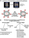
A, Principle of creating defects in the lipid bilayer. Top panel shows fatty acyl chain organization at the Tm temperature. The diagram was adapted from the website of Venable & Pastor (FDA/CBER). Lower panel shows release of drugs through localized defects in the membrane. B, Various strategies used for localized drug delivery.
FIGURE 5
FIGURE 5
Examples of external triggering modalities. A, Drug-loaded superparamagnetic iron oxide nanoparticles (SPIONs) encapsulated in liposome membrane (top panel). Exposure of these liposomes to alternative magnetic field results in local disruption of liposomes releasing SPIONS (step 1), and the drug is then released from the SPIONS (step 2). B, Timeline for development of thermosensitive liposomes. C and D, Chemical structures of designer lipids used for development of photo-activable liposomes: (a) Bis AzoPC, (b) Sorbyl PC, (c) o-nitrobenzyl conjugated lipid, (d) head-group polymerizable lipid, (e) DC8,9PC (1,2 bis(tricosa-10,12-diynoyl)-sn-glycero-3-phosphocholine), (f) principle of photopolymerization of DC8,9PC.
FIGURE 6
FIGURE 6
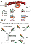
A, Targeting pathways of lipid-based nanoparticles to tumors. Schematic presentation of nanoparticles that passively or actively target tumors. These particles accumulate in the tumor area due to the leaky vasculature surrounding the tumors. However, targeted nanoparticles bind to the target tumor cells via ligand-receptor interactions and are internalized, whereas non-targeted nanoparticles are less efficient in interacting with tumor cells. B, Antibodies and antibody fragments used for targeting of lipid-based nanoparticles.
FIGURE 7
FIGURE 7
Scheme presenting steps in the conjugation of antibodies/targeting molecules to the liposomes. A, Post-insertion protocol. B, Antibody conjugation on the surface of liposomes.
FIGURE 8
FIGURE 8
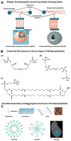
Bolaamphiphiles. A, Self-assembly of bolalipids into monolayers and comparison with bilayer forming lipids such as phosphatidylcholine. B, Chemical structures of various types of bolalipids: (a) bolaamphiphilic phosphocholine, (b) symmetrical bipolar phospholipid, and (c) unsymmetrical bipolar phospholipids. C, Possible assembly and aggregate structures of bolaamphiphiles.
FIGURE 9
FIGURE 9
Solid lipid nanoparticles (SLN) and nanostructured lipid carriers (NLC).

Similar articles

Cited by

References

    1. Wang AZ, Gu F, Zhang L, Chan JM, Radovic-Moreno A, Shaikh MR, Farokhzad OC. Biofunctionalized targeted nanoparticles for therapeutic applications. Expert Opin Biol Ther. 2008;8(8):1063–70. - PMC - PubMed
    1. Zhang L, Gu FX, Chan JM, Wang AZ, Langer RS, Farokhzad OC. Nanoparticles in medicine. therapeutic applications and developments. Clin Pharmacol Ther. 2008;83(5):761–9. - PubMed
    1. Alaouie AM, Sofou S. Liposomes with triggered content release for cancer therapy. J Biomed Nanotech. 2008;4(3):234–4.
    1. Allen TM, Cullis PR. Drug delivery systems: entering the mainstream. Science. 2004;303(5665):1818–22. - PubMed
    1. Alonso MJ. Nanomedicines for overcoming biological barriers. Biomed Pharmacother. 2004;58(3):168–72. - PubMed

Publication types