. 2010 Jul;114(1):271-80.
doi: 10.1111/j.1471-4159.2010.06749.x. Epub 2010 Apr 16.

Mitochondrial protection after traumatic brain injury by scavenging lipid peroxyl radicals

Affiliations

Mitochondrial protection after traumatic brain injury by scavenging lipid peroxyl radicals

Ayman G Mustafa et al. J Neurochem. 2010 Jul.

Abstract

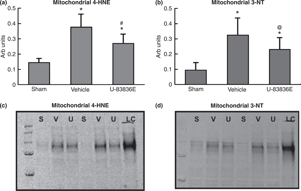
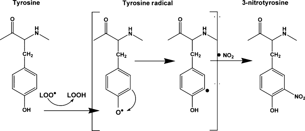
Mitochondrial dysfunction after traumatic brain injury (TBI) is manifested by increased levels of oxidative damage, loss of respiratory functions and diminished ability to buffer cytosolic calcium. This study investigated the detrimental effects of lipid peroxyl radicals (LOO(*)) and lipid peroxidation (LP) in brain mitochondria after TBI by examining the protective effects of U-83836E, a potent and selective scavenger of LOO(*) radicals. Male CF1 mice were subjected to severe controlled cortical impact TBI (CCI-TBI) and treated with either vehicle or U-83836E initiated i.v. at 15 min post-injury. Calcium (Ca(++)) buffering capacity and respiratory function were measured in isolated cortical mitochondrial samples taken from the ipsilateral hemisphere at 3 and 12 h post-TBI, respectively. In vehicle-treated injured mice, the cortical mitochondrial Ca(++) buffering capacity was reduced by 60% at 3 h post-injury (p < 0.001) and the respiratory control ratio was decreased by 27% at 12 h post-TBI, relative to sham, non-injured mice. U-83836E treatment significantly (p < 0.05) preserved Ca(++) buffering capacity and attenuated the reduction in respiratory control ratio values. Consistent with the functional effects of U-83836E being as a result of an attenuation of mitochondrial oxidative damage, the compound significantly (p < 0.001) reduced LP-generated 4-hydroxynonenal levels in both cortical homogenates and mitochondria at both 3 and 12 h post-TBI. Unexpectedly, U-83836E also reduced peroxynitrite-generated 3-nitrotyrosine in parallel with the reduction in 4-hydroxynonenal. The results demonstrate that LOO(*) radicals contribute to secondary brain mitochondrial dysfunction after TBI by propagating LP and protein nitrative damage in cellular and mitochondrial membranes.


Figures

Fig. 1
Fig. 1
U-83836 is a 2-methylaminochroman that contains the phenolic chroman ring structure of vitamin E (α-tocopherol, left side of diagram) which can donate an electron to a lipid peroxyl radical (LOO ) converting it to a lipid hydroperoxide (LOOH) which in turn can be converted to a harmless lipid alcohol (LOH) by the action of the antioxidant enzyme glutathione peroxidase (GSHPx). The chroman radical is a weak, non-oxidizing free radical that can be re-reduced back to the phenolic hydroxyl by ascorbate or glutathione (GSH) so that it can reduce a second LOO. On the right side of the diagram is shown the LOO scavenging mechanism for the bis-pyrrolo pyrimidine portion of U-83836E. Initially, a LOO binds to the 5 position of the pyrimidine ring as shown. This is hydrolyzed resulting in the formation of a lipid alcohol and a 5-hydroxy pyrimidine. This phenolic hydroxyl can react with an alkoxyl radical (LO) radical converting it to a LOOH which in turn can be converted by either GSHPx or phospholipid GSHPx to a LOH. The right hand reaction sequence represents a catalytic scavenging mechanism in which the antioxidant bis-pyrrolo pyrimidine moiety is able to repeatedly react with LOO and LO radicals. The dual peroxyl scavenging mechanism together with the ability of the chroman phenol to be re-reduced and the bis-pyrollo-pyrimdine to react catalytically makes U-83836 a highly effective and potent lipid peroxidation inhibitor. U-83836E = HCl salt of U-83836.
Fig. 2
Fig. 2
Attenuation of post-traumatic lipid peroxidation–derived 4-hydroxynonenal (4-HNE) (a) and protein nitration-related 3-nitrotyrosine (3-NT) (b) in cortical homogenates by U-83836E at 3 h following controlled cortical impact traumatic brain injury in male mice. Mice were administered 3 mg/kg of U-83836E i.v. 15 min post-injury and oxidative damage markers were measured 3 h following injury. The levels of 4-HNE and 3-NT were detected using western blotting, (c) and (d) respectively. Lanes were loaded with either sham (S), vehicle (V), U-83836E (U) or loading control (LC). Bars indicate group means ± SD. Statistical differences (one-way anova and Student-Neuman–Keuls post hoc test): *p < 0.001 versus sham, #p < 0.001 versus vehicle, n = 10/group.
Fig. 3
Fig. 3
Effects of U-83836E on cortical mitochondrial bioenergetics at 12 h following severe controlled cortical impact traumatic brain injury in male mice: (a). respiratory control ratio (RCR); (b). State III and IV respiratory rates. Animals were administered 3 mg/kg U-83836E i.v. 15 min post-injury and mitochondria isolated using the Ficoll density-gradient isolation technique. Mitochondrial oxygen consumption was measured using a Clark-type oxygen electrode. RCR was calculated as the ratio of oxygen consumption in the presence of ADP (state III) and after the addition of oligomycin (state IV). Values = mean ± sd. Statistical differences (one-way anova and Student-Neuman–Keuls post hoc test): *p < 0.05 versus sham, #p < 0.05 versus vehicle, n = 10/group.
Fig. 4
Fig. 4
Attenuation of lipid peroxidation–derived 4-hydroxynonenal (4-HNE) (a) and protein nitration-related 3-nitrotyrosine (3-NT) (b) in mitochondria by U-83836E at 12 h following controlled cortical impact traumatic brain injury in male mice. Animals were administered 3 mg/kg of U-83836E i.v. 15 min post-injury and oxidative markers were measured 12 h following injury. The levels of 4-HNE and 3-NT were detected using western blotting, (c) and (d) respectively. Lanes were loaded with either sham (S), vehicle (V), U-83836E (U) or loading control (LC). Values = mean ± SD. Statistical differences (one-way anova and Student-Neuman–Keuls post hoc test): *p < 0.001 versus sham, #p < 0.001 versus vehicle, @ p < 0.05 versus vehicle n = 10.
Fig. 5
Fig. 5
Effects of U-83836E on Ca++ buffering capacity in mitochondria at 3 h following controlled cortical impact traumatic brain injury in male mice. Mice were administered 3 mg/kg U-83836E i.v. 15 min post-injury and mitochondria were isolated using the Ficoll density-gradient isolation technique. Part (a) shows a quantification of mitochondrial Ca++ buffering capacity across experimental groups. The Ca++ buffering capacity was reduced in vehicle-treated injured group compared to sham (non-injured) group and U-83836E treatment ameliorated this posttraumatic reduction in mitochondrial Ca++ buffering capacity. Mitochondrial Ca++ buffering was evaluated in a thermostatically-controlled, continuously stirred cuvette incubated in a Shimadzu RF-5301 spectrofluorimeter set at 37°C. Levels of extra-mitochondrial calcium were measured using Ca++-sensitive indicator calcium green 5 N (CaG5N), a fluorescent calcium-sensitive indicator. Malate and pyruvate (M/P) and ADP were loaded sequentially over 2 min followed by oligomycin (O) at 3 min. Two minutes after adding oligomycin, calcium (32 nmol/L) infusion started at a rate of 0.5µL/min. Part (b) is a typical trace that shows the differences in mitochondrial Ca++ uptake in sham (non-injured), vehicle-treated injured and U-83836E-treated injured mitochondria. Values = mean ± sd. Statistical differences (one-way anova and Student-Neuman–Keuls post hoc test): *p < 0.01 versus sham, #p < 0.05 versus vehicle, n = 4.
Fig. 6
Fig. 6
Mechanistic hypothesis of the contribution of lipid peroxyl radicals to peroxynitrite-mediated oxidative damage following traumatic brain injury (see Discussion).

Similar articles

Cited by

References

    1. Alvarez B, Radi R. Peroxynitrite reactivity with amino acids and proteins. Amino Acids. 2003;25:295–311. - PubMed
    1. Beckman JS, Koppenol WH. Nitric oxide, superoxide, and peroxynitrite: the good, the bad, ugly. Am. J. Physiol. 1996;271:C1424–C1437. - PubMed
    1. Brown MR, Sullivan PG, Dorenbos KA, Modafferi EA, Geddes JW, Steward O. Nitrogen disruption of synaptoneurosomes: an alternative method to isolate brain mitochondria. J. Neurosci. Methods. 2004;137:299–303. - PubMed
    1. Brustovetsky N, Brustovetsky T, Jemmerson R, Dubinsky JM. Calcium-induced cytochrome c release from CNS mitochondria is associated with the permeability transition and rupture of the outer membrane. J. Neurochem. 2002;80:207–218. - PubMed
    1. Budd SL, Nicholls DG. A reevaluation of the role of mitochondria in neuronal Ca2+ homeostasis. J. Neurochem. 1996;66:403–411. - PubMed

MeSH terms