. 2010 Jul;8(7):1584-93.
doi: 10.1111/j.1538-7836.2010.03883.x. Epub 2010 Apr 16.

PECAM-1 functions as a negative regulator of laminin-induced platelet activation

Affiliations

PECAM-1 functions as a negative regulator of laminin-induced platelet activation

J Crockett et al. J Thromb Haemost. 2010 Jul.

Abstract

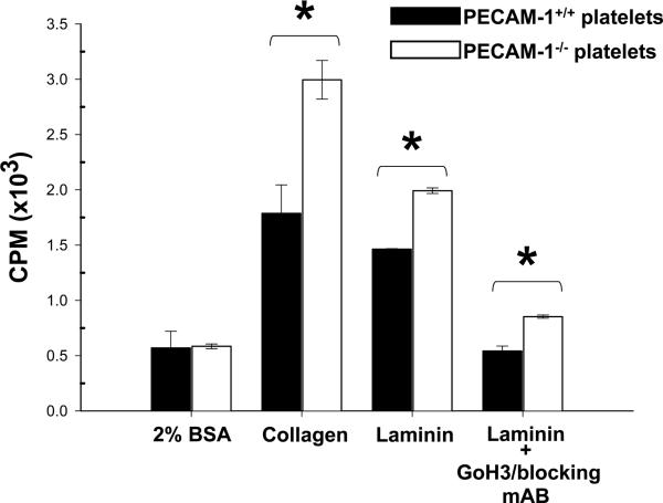
Summary background: Interaction of resting platelets with exposed components of the subendothelial matrix is an important early activating event that takes place at sites of vascular injury. Platelet responses to collagen are mediated by integrin alpha(2)beta(1) and the glycoprotein (GP)VI-Fc receptor (FcR) gamma-chain complex, whereas platelet activation by laminin is mediated by the related integrin, alpha(6)beta(1), and similarly requires signaling through GPVI-FcR gamma-chain.

Objective: Because the cell adhesion and signaling receptor PECAM-1 has previously been shown to dampen collagen-induced platelet activation, we sought to determine whether PECAM-1 might similarly regulate platelet activation by laminin.

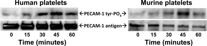
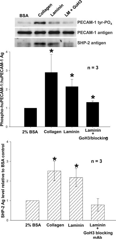
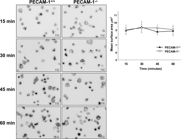
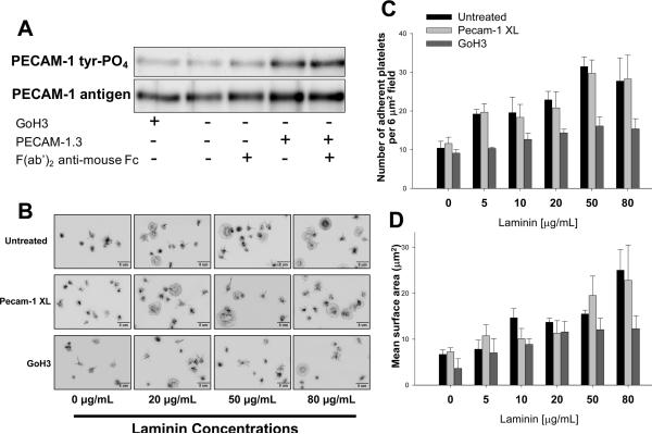
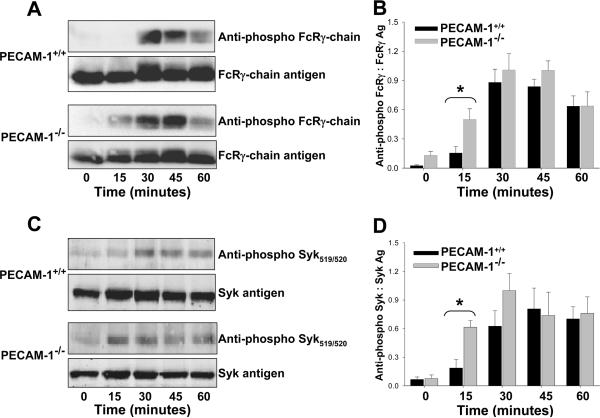
Methods/results: We found that PECAM-1 became tyrosine phosphorylated on its cytoplasmic immunoreceptor tyrosine-based inhibitory motifs following adhesion of either human or murine platelets to immobilized laminin. Whereas the presence or absence of PECAM-1 had no effect on either the rate or extent of platelet adhesion or spreading on laminin, PECAM-1 inhibited laminin-induced phosphorylation of GPVI-FcR gamma-chain immunoreceptor tyrosine-based activation motifs (ITAMs) and activation of its downstream effector, Syk kinase, and suppressed granule secretion.

Conclusions: Taken together, these data are consistent with previous findings in platelets and other blood and vascular cells that PECAM-1 functions by modulating ITAM-mediated signaling pathways that amplify cellular activation.


Figures

Figure 1
Figure 1. Human platelets adhere to immobilized laminin and collagen, but spread with different morphology
(A) Visualization of platelets bound to immobilized matrix proteins. Washed human platelets resuspended at 1 × 10⁸/mL in 2 mM MgCl2 and 0.5 mM CaCl2 were seeded on surfaces pre-coated with either human placental laminin 511 or soluble calf-skin collagen. After 30 minutes at 37°C, unbound platelets were removed, adherent platelets fixed with 3% paraformaldehyde, and visualized using Hoffman modulation contrast. Results shown are representative of three separate experiments. Note that platelets bind well to immobilized laminin, but do not spread as well as they do on immobilized collagen. (B) Quantitation of platelet adhesion to immobilized matrix proteins. Washed human platelets were loaded with Calcein AM and seeded onto microtiter wells that had been pre-coated with the indicated concentrations of human laminin or collagen. After a 60 minute incubation at 37°C, the percentage of adherent platelets was calculated by taking fluorescence measurements both before and after removal of unbound platelets. Data shown represents mean values ± SEM from three independent experiments performed using triplicate wells.
Figure 2
Figure 2. PECAM-1 becomes tyrosine phosphorylated following adhesion of human and mouse platelets to immobilized laminin
Washed platelets were resuspended at a concentration of 4 × 10⁸/mL in Tyrodes buffer containing 2 mM MgCl2 and 0.5 mM CaCl2 and added to culture plates that had been pre-coated with 50 μg/mL of laminin 511. Platelets were allowed to settle, bind, and spread at 37°C for the indicated times, and then detergent lysed and subjected to immunoprecipitation analysis using mAbs PECAM-1.3 for human platelets and 390 for murine platelets. Western blots were developed using the anti-phosphotyrosine mAb PY20 (top panels) and anti-PECAM-1 polyclonal antibodies SEW32-34 (human) and M-20 (mouse) to visualize antigen loading. Maximal phosphorylation of PECAM-1 at the 45 minute time point reflects the time needed for platelets to settle, make contact with the immobilized laminin, and activate the platelets. The result shown is representative of three separate experiments.
Figure 3
Figure 3. Tyrosine-phosphorylated PECAM-1 recruits SHP-2 following adhesion to immobilized laminin
Top panel: Washed human platelets were added to laminin-coated tissue culture plates as described in the legend for Figure 2 and incubated for 30-45 minutes at 37°C. Wells coated with 1 mg/ml BSA or 50 μg/mL collagen served as negative and positive controls, respectively. Platelets were then lysed and subjected to immunoprecipitation/western blot analysis as in Figure 2. SHP-2 was detected in the co-immunoprecipitates using polyclonal antibody C-18. The result shown is representative of three individual experiments. Middle and bottom panels: Quantitative analysis of PECAM-1 tyrosine phosphorylation and SHP-2 binding following platelet adhesion to immobilized laminin. Band intensity was determined using a Kodak Molecular Imaging Densitometry System. PECAM-1 tyrosine phosphorylation levels are expressed as the ratio of PY20:PECAM-1 antigen. Data were normalized to values obtained from resting platelets adherent to BSA. Data from three independent experiments were analyzed by the Wilcoxon rank sum test and represented as mean ± SEM. *P < 0.05. Similar results were obtained for murine platelets (not shown).
Figure 4
Figure 4. Rate and extent of spreading of wild-type versus PECAM-1–deficient platelets on immobilized laminin
Washed platelets isolated from the anticoagulated whole blood of wild-type or PECAM-1–deficient mice were seeded on laminin-coated coverslips in the presence of 2 mM Mg++ and incubated for 15, 30, 45 or 60 minutes at 37°C. Adherent platelets were fixed, permeabilized, stained with TRITC-labeled phalloidin, and visualized by fluorescence microscopy. Left panels – representative images taken over a 60 minute period. Upper right panel - Quantitation of platelet spreading from eight randomly chosen fields (~200 platelets/field) using Metamorph software. Data represent the mean ± SEM from three independent experiments. Statistical significance was determined using the Student t-test for unpaired samples.
Figure 5
Figure 5. PECAM-1 engagement-induced inhibitory signaling does not affect platelet adhesion or spreading on immobilized laminin
(A) 3 × 10⁸ human platelets/ml were incubated with 5 μg/mL mAb PECAM-1.3 for 10 minutes in the presence or absence of F(ab')2 fragments of anti-mouse IgG Fc and subjected to immunoprecipitation/western blot analysis. GoH3, a specific mAb for the integrin α6 subunit, was used as a negative control. Note that binding and cross-linking of mAb PECAM-1.3 on the platelet surface was able to induce tyrosine phosphorylation of PECAM-1. (B) Effect of antibody-induced inhibitory signaling on platelet spreading on immobilized laminin. Resting human platelets were treated with GoH3 or mAb PECAM-1.3 and cross-linked (Pecam-1 XL) as described in panel A and the platelets added to culture dishes that had been the indicated concentration of laminin. After 30 minutes at 37°C, unbound platelets were removed and the remaining bound platelets were fixed, stained, and analyzed. Panels C and D - The number and mean surface area of adherent platelets from three independent experiments were quantitated from 8 randomly chosen fields of 50 – 300 platelets each, analyzed using Metamorph software, and expressed as the mean ± SEM. Note that although PECAM-1.3 was effective in initiating inhibitory signaling (panel A), this had no effect on platelet adhesion or spreading.
Figure 6
Figure 6. PECAM-1 regulates laminin-induced granule secretion
Washed platelets obtained from age- and sex-matched wild-type or PECAM-1-deficient mice were labeled with 14C-serotonin and added in triplicate to wells pre-coated with either 2% BSA, 50 μg/mL of collagen, or 50 μg/mL of laminin. In some cases, platelets were preincubated with 5 μg/mL of GoH3 10 minutes before seeding onto immobilized laminin. After 60 minutes at 37°C, the amount of 14C-serotonin that had been released into the culture media was determined by scintillation counting and expressed as the mean ± SEM. Data shown is representative of two independent experiments. * P < 0.001.
Figure 7
Figure 7. Early activation of the GPVI /FcRγ-chain→Syk platelet activation pathway in PECAM-1-deficient platelets
Murine platelets were added to laminin-coated microtiter wells for the indicated times, lysed, and subjected to western blot analysis using the indicated antibodies. Panels A and B - PECAM-1 delays laminin-induced FcRγ-chain phosphorylation. A representative immunoblot is shown in (A), and cumulative quantitative data derived from three independent experiments is shown in (B). Panels C and D - PECAM-1 affects the kinetics of laminin-induced Syk phosphorylation. A representative immunoblot is shown in (C), with cumulative quantitative data derived from three independent experiments is shown in (D). Note that PECAM-1 appears to set a higher threshold for platelet activation, as both the FcRγ chain and Syk become phosphorylated earlier in PECAM-1-deficient platelets.

Similar articles

Cited by

References

    1. Scheele S, Nystrom A, Durbeej M, Talts JF, Ekblom M, Ekblom P. Laminin isoforms in development and disease. J Mol Med. 2007;85:825–36. - PubMed
    1. Jackson SP. The growing complexity of platelet aggregation. Blood. 2007;109:5087–95. - PubMed
    1. Newman PJ, Newman DK. Platelets and the Vessel Wall. 7th Edition 2008. pp. 1378–98.
    1. Watson SP, Auger JM, McCarty OJ, Pearce AC. GPVI and integrin αIIb β3 signaling in platelets. J Thromb Haemost. 2005;3:1752–62. - PubMed
    1. Newton-Nash DK, Newman PJ. A new role for PECAM-1 (CD31): Inhibition of TCR-mediated signal transduction. J Immunol. 1999;163:682–8. - PubMed

Publication types

Substances