. 2010 Apr 19:9:100.
doi: 10.1186/1475-2875-9-100.

The kinetics of antibody binding to Plasmodium falciparum VAR2CSA PfEMP1 antigen and modelling of PfEMP1 antigen packing on the membrane knobs

Affiliations

The kinetics of antibody binding to Plasmodium falciparum VAR2CSA PfEMP1 antigen and modelling of PfEMP1 antigen packing on the membrane knobs

Lars M Joergensen et al. Malar J. .

Abstract

Background: Infected humans make protective antibody responses to the PfEMP1 adhesion antigens exported by Plasmodium falciparum parasites to the erythrocyte membrane, but little is known about the kinetics of this antibody-receptor binding reaction or how the topology of PfEMP1 on the parasitized erythrocyte membrane influences antibody association with, and dissociation from, its antigenic target.

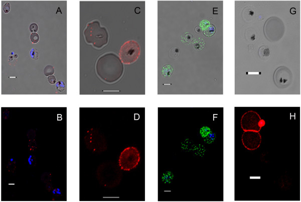
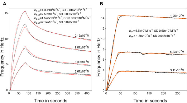
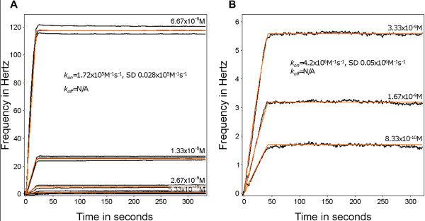
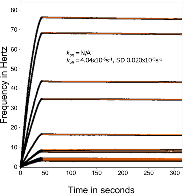
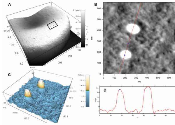
Methods: A Quartz Crystal Microbalance biosensor was used to measure the association and dissociation kinetics of VAR2CSA PfEMP1 binding to human monoclonal antibodies. Immuno-fluorescence microscopy was used to visualize antibody-mediated adhesion between the surfaces of live infected erythrocytes and atomic force microscopy was used to obtain higher resolution images of the membrane knobs on the infected erythrocyte to estimate knob surface areas and model VAR2CSA packing density on the knob.

Results: Kinetic analysis indicates that antibody dissociation from the VAR2CSA PfEMP1 antigen is extremely slow when there is a high avidity interaction. High avidity binding to PfEMP1 antigens on the surface of P. falciparum-infected erythrocytes in turn requires bivalent cross-linking of epitopes positioned within the distance that can be bridged by antibody. Calculations of the surface area of the knobs and the possible densities of PfEMP1 packing on the knobs indicate that high-avidity cross-linking antibody reactions are constrained by the architecture of the knobs and the large size of PfEMP1 molecules.

Conclusions: High avidity is required to achieve the strongest binding to VAR2CSA PfEMP1, but the structures that display PfEMP1 also tend to inhibit cross-linking between PfEMP1 antigens, by holding many binding epitopes at distances beyond the 15-18 nm sweep radius of an antibody. The large size of PfEMP1 will also constrain intra-knob cross-linking interactions. This analysis indicates that effective vaccines targeting the parasite's vulnerable adhesion receptors should primarily induce strongly adhering, high avidity antibodies whose association rate constant is less important than their dissociation rate constant.


Figures

Figure 1
Figure 1
Patient plasma IgG and anti-PfEMP1 IgG binding to IE. IE incubated with goat anti-human IgG conjugated to Alexa®568 (red) or goat anti-rabbit IgG Alexa®488 (green). A. Antibody selected IE (3D7) incubated with 1:50 diluted Tanzanian child's acute-phase malaria serum. The DIC images of IE show DAPI stained parasite nuclei (blue) and spots of membrane-bound IgG (Alexa®568). B. Fluorescence image of Figure 1A. C. Higher magnification DIC image of live IE (3D7) and uninfected erythrocytes, incubated with 1:100 diluted convalescent plasma from the same child. D. Fluorescence image of Figure 1C. E. DIC images of 3D7, incubated with rabbit antiserum against a recombinant Var4 PfEMP1, detected with goat anti-rabbit IgG (Alexa®488). F. Fluorescence image of Figure 1E. G. DIC and DAPI (blue) fluorescence image of immune serum-agglutinated and unagglutinated erythrocytes. IE (FCR3) selected by panning on Dynabeads coated with IgG from a pool of semi-immune Tanzanian children's sera (note DAPI-staining merozoite invading the upper IE). H. Agglutination of two IE by serum used in Figures 1C and 1D, detected with goat anti-human IgG Alexa®568. Two infected and one uninfected erythrocytes are unagglutinated and largely unstained. The invading merozoite is stained by serum IgG. Live trophozoites often do not take up DAPI but such IE can be identified by pigment or serum staining (Figure 1E). Scale bars 5 μm.
Figure 2
Figure 2
Association and dissociation of a weakly binding antibody to VAR2CSA DBL5ε. The frequency change (Hz) as a function of time (s), shows both the association and dissociation phase of the PAM 4.7 antibody binding to the DBL5ε domain of the VAR2CSA PfEMP1. Black lines indicate data; red lines indicate fitted curves (see Methods) and the calculated rate constants are given on the sensorgrams. A. PAM 4.7, in flow, reacting with VAR2CSA DBL5ε immobilized on a polystyrene surface, tested at the four concentrations shown. B. PAM 4.7, in flow, reacting with VAR2CSA DBL5ε, immobilized on a carboxyl surface and tested at three concentrations with a lower density of surface-immobilized antigen.
Figure 3
Figure 3
Association and dissociation of a strongly-binding antibody with different surface densities of VAR2CSA DBL5ε. The frequency shift (Hz) is shown as a function of time (s). Black lines represent data from different reactions, with the indicated concentrations of PAM 3.10 binding to VAR2CSA DBL5ε immobilized on a carboxyl surface. Red lines indicate the fitted curves, as described in the Methods. The rate constants calculated are given on the sensorgram. A. PAM 3.10, in flow, against a high density surface. B. PAM 3.10, in flow, against a low density surface. See Additional file 1 for surface antigen density calculations.
Figure 4
Figure 4
The interaction of monovalent Fab fragment of PAM 3.10 with VAR2CSA DBL5ε. Association and dissociation of the Fab fragments of PAM 3.10 to a high density of VAR2CSA DBL5ε on the sensor chip surface. The frequency change is shown as a function of time and the black lines show the data from binding experiments with different concentrations of the PAM 3.10 Fab fragment to the surface-bound protein. The red lines show the curve fit for the dissociation phase only. The dissociation rate constant calculated is given on the sensorgram.
Figure 5
Figure 5
AFM derived measurement of the dimensions of the knob structures on the IE membrane. Force microscopy was carried out on unfixed, air dried IE. A. AFM tapping mode image of a single trophozoite-IE (FCR3). The boxed insert indicates the two knob structures analysed at higher resolution. B. Higher resolution scan data from the two knobs boxed in Figure 5A, bisected by the red sectioning line with the knob centre and a selected circumferential point indicated by the blue points. C. Two-colour enhanced three dimensional imaging of tapping-mode scan data. D. Topology measurements of the surface bisected by the red line shown in Figure 5B.

Similar articles

Cited by

References

    1. Bull PC, Lowe BS, Kortok M, Molyneux CS, Newbold CI, Marsh K. Parasite antigens on the infected red cell are targets for naturally acquired immunity to malaria. Nat Med. 1998;4:358–360. doi: 10.1038/nm0398-358. - DOI - PMC - PubMed
    1. Dodoo D, Staalsoe T, Giha H, Kurtzhals JAL, Akanmori BD, Koram K, Dunyo S, Nkrumah FK, Hviid L, Theander TG. Antibodies to variant antigens on the surfaces of infected erythrocytes are associated with protection from malaria in Ghanaian children. Infect Immun. 2001;69:3713–3718. doi: 10.1128/IAI.69.6.3713-3718.2001. - DOI - PMC - PubMed
    1. Giha HA, Staalsoe T, Dodoo D, Roper C, Satti GM, Arnot DE, Hviid L, Theander TG. Antibodies to variable Plasmodium falciparum-infected erythrocyte surface antigens are associated with protection from novel malaria infections. Immunol Lett. 2000;71:117–126. doi: 10.1016/S0165-2478(99)00173-X. - DOI - PubMed
    1. Zhang J, Su XD, O'Shea SJ. Antibody/antigen affinity behavior in liquid environment with electrical impedance analysis of quartz crystal microbalances. Biophys Chem. 2002;99:31–41. doi: 10.1016/S0301-4622(02)00109-6. - DOI - PubMed
    1. Steckbeck JD, Orlov I, Chow A, Grieser H, Miller K, Bruno J, Robinson JE, Montelaro RC, Cole KS. Kinetic rates of antibody binding correlate with neutralization sensitivity of variant simian immunodeficiency virus strains. J Virol. 2005;79:12311–12320. doi: 10.1128/JVI.79.19.12311-12320.2005. - DOI - PMC - PubMed

MeSH terms

Substances