

Increased expression of urokinase plasminogen activator and its cognate receptor in human seminomas
- PMID: 20403162
- PMCID: PMC2885360
- DOI: 10.1186/1471-2407-10-151
Increased expression of urokinase plasminogen activator and its cognate receptor in human seminomas
Abstract
Background: The urokinase plasminogen activating system (uPAS) is implicated in neoplastic progression and high tissue levels of uPAS components correlate with a poor prognosis in different human cancers. Despite that, relative few studies are available on the expression and function of the uPAS components in human seminomas. In the present study we characterized the expression of the urokinase plasminogen activator (uPA), its cognate receptor (uPAR) and the uPA inhibitors PAI-1 and PAI-2 in normal human testis and seminomas.
Methods: The expression of the above genes was evaluated by means of quantitative RT-PCR, western blot, zymographic analysis and immunohistochemistry.
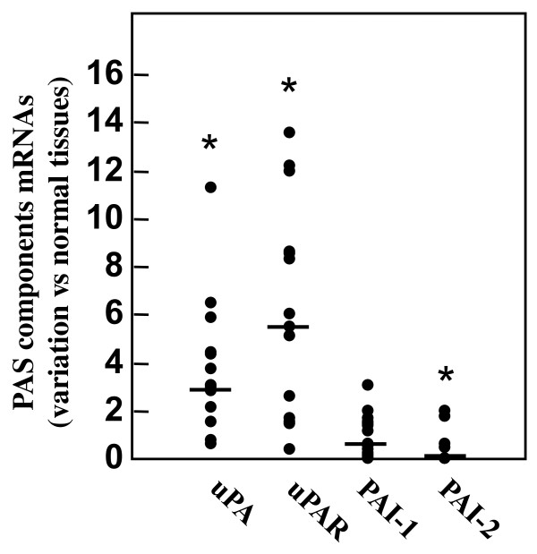
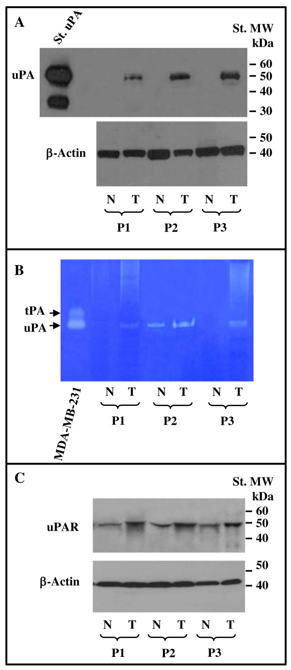
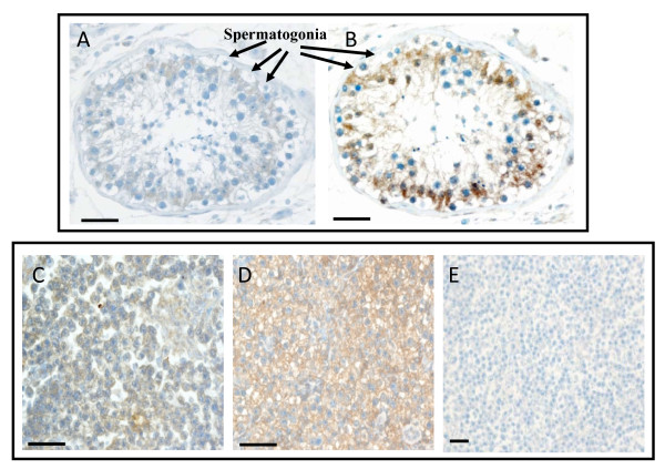
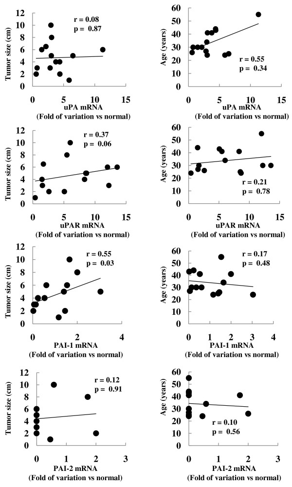
Results: Quantitative RT-PCR analysis of 14 seminomas demonstrated that uPA and uPAR mRNAs were, with respect to control tissues, increased in tumor tissues by 3.80 +/- 0.74 (p < 0.01) and 6.25 +/- 1.18 (p < 0.01) fold, respectively. On the other hand, PAI-1 mRNA level was unchanged (1.02 +/- 0.24 fold), while that of PAI-2 was significantly reduced to 0.34 +/- 0.18 (p < 0.01) fold. Western blot experiments performed with protein extracts of three seminomas and normal tissues from the same patients showed that uPA protein levels were low or undetectable in normal tissues and induced in tumor tissues. On the same samples, zymographic analysis demonstrated increased uPA activity in tumor tissue extracts. Western blot experiments showed that also the uPAR protein was increased in tumor tissues by 1.83 +/- 0.15 fold (p < 0.01). The increased expression of uPA and uPAR was further confirmed by immunohistochemical staining performed in 10 seminomas and autologous uninvolved peritumoral tissues. Finally, variation in the mRNA level of PAI-1 significantly correlated with tumor size.
Conclusions: We demonstrated the increased expression of uPA and uPAR in human seminomas with respect to normal testis tissues, which may be relevant in testicular cancer progression.
Figures
Similar articles
-
High expression of the urokinase plasminogen activator and its cognate receptor associates with advanced stages and reduced disease-free interval in papillary thyroid carcinoma.J Clin Endocrinol Metab. 2011 Feb;96(2):504-8. doi: 10.1210/jc.2010-1688. Epub 2010 Nov 24. J Clin Endocrinol Metab. 2011. PMID: 21106716
-
Combined mRNA expression levels of members of the urokinase plasminogen activator (uPA) system correlate with disease-associated survival of soft-tissue sarcoma patients.BMC Cancer. 2011 Jun 25;11:273. doi: 10.1186/1471-2407-11-273. BMC Cancer. 2011. PMID: 21702998 Free PMC article.
-
Significant down-regulation of the plasminogen activator inhibitor 1 mRNA in pancreatic cancer.Pancreas. 2008 Mar;36(2):173-7. doi: 10.1097/MPA.0b013e31815ac538. Pancreas. 2008. PMID: 18376309
-
[The urokinase-type plasminogen activator system and its role in tumor progression].Biomed Khim. 2018 Nov;64(6):472-486. doi: 10.18097/PBMC20186406472. Biomed Khim. 2018. PMID: 30632975 Review. Russian.
-
The Urokinase Plasminogen Activation System in Pancreatic Cancer: Prospective Diagnostic and Therapeutic Targets.Biomolecules. 2022 Jan 18;12(2):152. doi: 10.3390/biom12020152. Biomolecules. 2022. PMID: 35204653 Free PMC article. Review.
Cited by
-
Prognostic value of intratumoral carbonic anhydrase IX expression in testicular germ cell tumors.Oncol Lett. 2017 Apr;13(4):2177-2185. doi: 10.3892/ol.2017.5745. Epub 2017 Feb 14. Oncol Lett. 2017. PMID: 28454378 Free PMC article.
-
Tazarotene-Induced Gene 1 (TIG1) Interacts with Serine Protease Inhibitor Kazal-Type 2 (SPINK2) to Inhibit Cellular Invasion of Testicular Carcinoma Cells.Biomed Res Int. 2019 Nov 25;2019:6171065. doi: 10.1155/2019/6171065. eCollection 2019. Biomed Res Int. 2019. PMID: 31886233 Free PMC article.
-
Prognostic value of programmed-death-1 receptor (PD-1) and its ligand 1 (PD-L1) in testicular germ cell tumors.Ann Oncol. 2016 Feb;27(2):300-5. doi: 10.1093/annonc/mdv574. Epub 2015 Nov 23. Ann Oncol. 2016. PMID: 26598537 Free PMC article.
-
A Role of the TEX101 Interactome in the Common Aetiology Behind Male Subfertility and Testicular Germ Cell Tumor.Front Oncol. 2022 Jun 14;12:892043. doi: 10.3389/fonc.2022.892043. eCollection 2022. Front Oncol. 2022. PMID: 35774118 Free PMC article. Review.
-
Increased levels of XPA might be the basis of cisplatin resistance in germ cell tumours.BMC Cancer. 2020 Jan 6;20(1):17. doi: 10.1186/s12885-019-6496-1. BMC Cancer. 2020. PMID: 31906898 Free PMC article.
References
-
- Bunge G, Bradbury JT. An early human seminoma. JAMA. 1965;193:960–962. - PubMed
MeSH terms
Substances
LinkOut - more resources
Full Text Sources
Medical
Miscellaneous
