Comparative Study
. 2010 Apr 20:10:39.
doi: 10.1186/1471-230X-10-39.

Role of myosin light chain kinase in intestinal epithelial barrier defects in a rat model of bowel obstruction

Affiliations
Comparative Study

Role of myosin light chain kinase in intestinal epithelial barrier defects in a rat model of bowel obstruction

Chi-Chin Wu et al. BMC Gastroenterol. .

Abstract

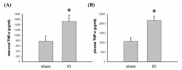
Background: Bowel obstruction is a common cause of abdominal emergency, since the patients are at increased risk of septicemia resulting in high mortality rate. While the compartmentalized changes in enteric microfloral population and augmentation of bacterial translocation (BT) have already been reported using experimental obstruction models, alterations in epithelial permeability of the obstructed guts has not been studied in detail. Myosin light chain kinase (MLCK) is actively involved in the contraction of epithelial perijunctional actinomyosin ring and thereby increases paracellular permeability. In the current study we attempt to investigate the role of MLCK in epithelial barrier defects using a rat model of simple mechanical obstruction.

Methods: Wistar rats received intraperitoneal injection of ML-7 (a MLCK inhibitor) or vehicle at 24, 12 and 1 hrs before and 12 hrs after intestinal obstruction (IO). The distal small intestine was obstructed with a single ligature placed 10 cm proximal to the ileocecal junction in IO rats for 24 hrs. Sham-operated rats served as controls.

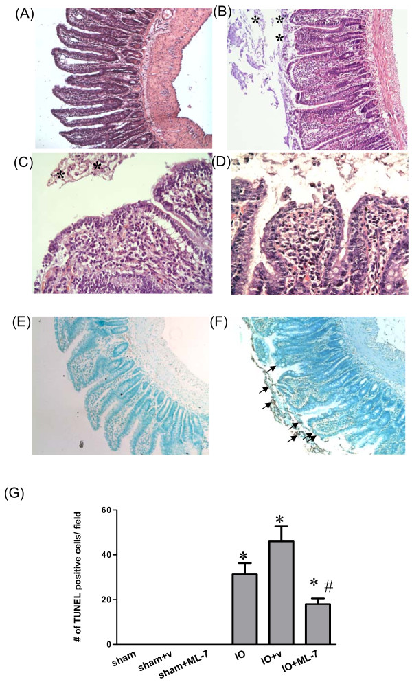
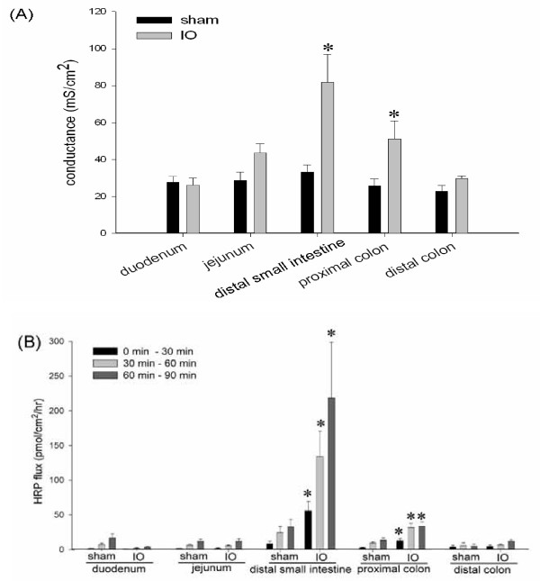
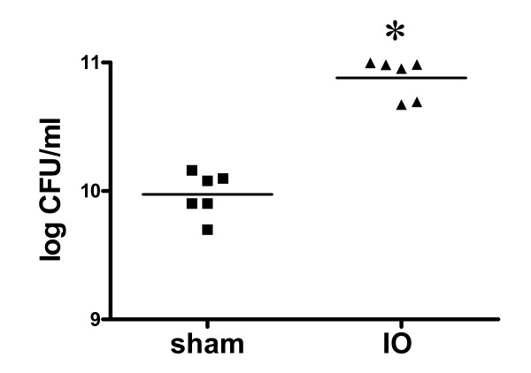
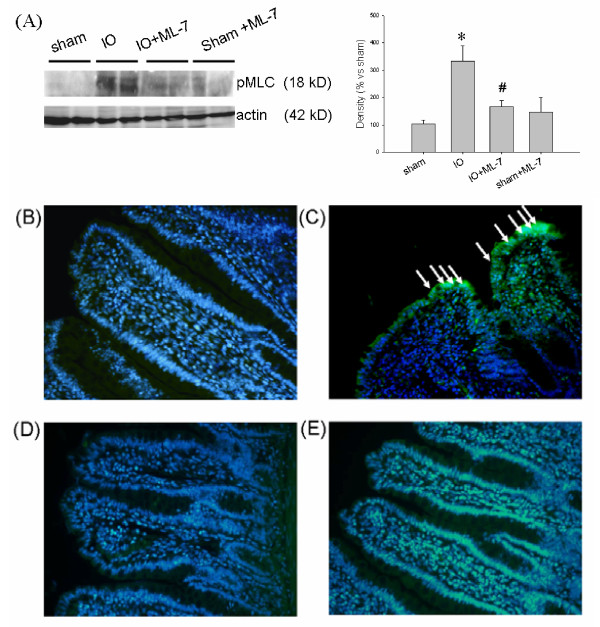
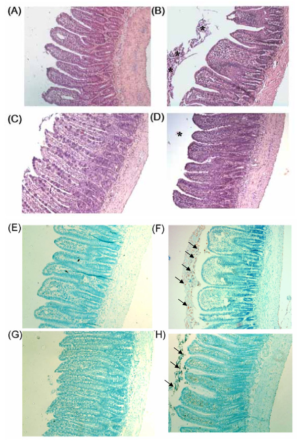
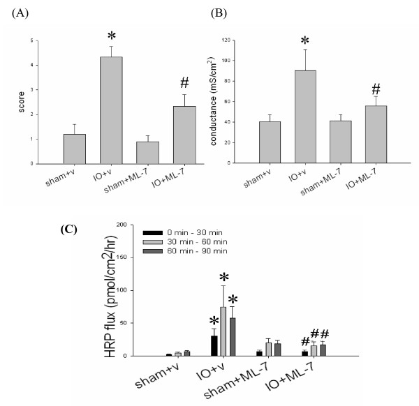
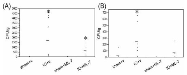
Results: Mucosal injury, such as villous blunting and increased crypt/villus ratio, was observed in the distal small intestine of IO rats. Despite massive enterocyte shedding, intestinal villi were covered with a contiguous epithelial layer without cell apoptosis. Increased transmural macromolecular flux was noticed in the distal small intestine and the proximal colon after IO. The bacterial colony forming units in the spleen and liver of IO rats were significantly higher than those of sham controls. Addition of ML-7 ameliorated the IO-triggered epithelial MLC phosphorylation, mucosal injury and macromolecular flux, but not the level of BT.

Conclusions: The results suggest that IO-induced premature enterocytic sloughing and enhanced paracellular antigenic flux were mediated by epithelial MLCK activation. In addition, enteric bacteria may undergo transcytotic routes other than paracellular paths to cross the epithelium.


Figures

Figure 1
Figure 1
Histological images of the distal small intestine of sham and IO rats. (Panels A-D) Sections of intestinal tissues were stained with H & E. (Panels E and F) The apoptotic level of intestinal tissues was examined by TUNEL assay. (A) Normal villous structure was seen in the distal small intestine of sham rats. (B) Following IO, villi were edematous and blunted. Severe shedding of epithelial cells was observed in the intestinal lumen of IO rats (asterisk). (C) Higher magnification of the epithelial layer in the distal small intestine of IO rats showing epithelial shedding without villous denudation. (D) A positive control displaying epithelial discontinuity in an ischemic gut model. (E) No sign of TUNEL(+) cells was observed in the intestine of sham controls. (F) A high number of apoptotic cells was seen in the gut lumen following IO (arrows). (G) Quantification of TUNEL(+) cells. *p < 0.05 vs. respective sham groups. #p < 0.05 vs. IO+v rats. Panels A, B, E and F: 100× magnification. Panels C and D: 400× magnification.
Figure 2
Figure 2
Enhanced mucosal and plasma TNFα levels following IO. (A) The TNFα levels in the mucosa of the distal small intestines in sham and IO rats. (B) The plasma TNFα concentration in sham and IO rats. n = 5/group. *p < 0.05 vs. sham rats.
Figure 3
Figure 3
IO induced epithelial permeability rise in the distal small intestine and proximal colon. Various bowel segments in sham and IO rats were mounted on Ussing chambers for the measurement of electrical conductance (Panel A) and mucosal-to-serosal flux of HRP (Panel B). The rate of HPR flux was determined at different time points after luminal addition of the probe: 0 to 30, 30 to 60, and 60 to 90 min. n = 5-7/group. *p < 0.05 vs. respective sham groups.
Figure 4
Figure 4
Bacterial overgrowth in the small intestinal lumen in IO rats. A tenfold increase of bacterial colony forming units (CFU) was seen in the intestinal lavage in IO rats compared to sham controls. Each data point in the figure represents the value from one animal, and the mean values of bacterial counts are shown as bars. n = 6/group. *p < 0.05 vs. sham rats.
Figure 5
Figure 5
Increased phosphorylation of myosin light chain in intestinal epithelium in IO rats was inhibited by treatment with ML-7. (A) Western blots showing that IO augmented the mucosal levels of phosphorylated MLC (pMLC), which was attenuated by ML-7. n = 3-4/group. *p < 0.05 vs. respective sham rats. #p < 0.05 vs. IO rats. Representative images of pMLC staining in intestinal tissues of (B) sham, (C) IO, (D) sham+ML-7, and (E) IO+ML-7 rats. The staining of pMLC (green color) was localized to the villous surface (arrows) in IO rats, whereas no staining was seen in sham controls. The cell nuclei were shown in blue color. Addition of ML-7 prevented the IO-induced phosphorylation of epithelial MLC. Images: 200× magnification.
Figure 6
Figure 6
Histological images of the distal small intestine of sham and IO rats treated with vehicle or ML-7. (Panels A to D) Sections of intestinal tissues were stained with H & E. (Panels E to H) The apoptotic level of intestinal tissues was determined by TUNEL assay. All photomicrographs were taken at 100× magnification. Representative images shown were intestinal tissues of four groups of rats: (A, E) sham+v, (B, F) IO+v, (C, G) sham+ML-7, and (D, H) IO+ML-7. The IO-induced villous blunting, epithelial shedding (asterisk) and cell apoptosis (arrows) were partially prevented by ML-7, but not by vehicle.
Figure 7
Figure 7
Administration of ML-7 reduced the histopathological score, and inhibited the epithelial permeability rise in IO rats. Intestinal tissues of sham and IO rats treated with vehicle (v) or ML-7 were graded for histological damage (A), or mounted on Ussing chambers for the measurement of electrical conductance (B) and mucosal-to-serosal HRP fluxes (C). n = 5-7/group. *p < 0.05 vs. sham rats. #p < 0.05 vs. IO+v rats.
Figure 8
Figure 8
IO-induced augmentation of BT was not prevented by ML-7. (A) Spleen and (B) liver tissues of sham and IO rats treated with vehicle (v) or ML-7 were examined for bacterial growth. Each dot represents the bacterial CFU in the visceral organ of an individual rat. The line indicates the mean for each group. n = 5-7/group. *p < 0.05 vs. respective sham rats.

Similar articles

Cited by

References

    1. Cappell MS, Batke M. Mechanical obstruction of the small bowel and colon. Med Clin North Am. 2008;92:575–597. doi: 10.1016/j.mcna.2008.01.003. - DOI - PubMed
    1. Diaz JJ, Bokhari F, Mowery NT, Acosta JA, Block EF, Bromberg WJ, Collier BR, Cullinane DC, Dwyer KM, Griffen MM, Mayberry JC, Jerome R. Guidelines for management of small bowel obstruction. J Trauma. 2008;64:1651–1664. doi: 10.1097/TA.0b013e31816f709e. - DOI - PubMed
    1. Blair SL, Chu DZ, Schwarz RE. Outcome of palliative operations for malignant bowel obstruction in patients with peritoneal carcinomatosis from nongynecological cancer. Ann Surg Oncol. 2001;8:632–637. doi: 10.1007/s10434-001-0632-1. - DOI - PubMed
    1. Sagar PM, MacFie J, Sedman P, May J, Mancey-Jones B, Johnstone D. Intestinal obstruction promotes gut translocation of bacteria. Dis Colon Rectum. 1995;38:640–644. doi: 10.1007/BF02054126. - DOI - PubMed
    1. Shiomi H, Shimizu T, Endo Y, Murata S, Kurumi Y, Uji Y, Tani T. Relations among circulating monocytes, dendritic cells, and bacterial translocation in patients with intestinal obstruction. World J Surg. 2007;31:1806–1812. doi: 10.1007/s00268-007-9110-7. - DOI - PubMed

Publication types